Figure 10. Immunolabeling for
Mn-superoxide dismutase (MnSOD) in human retinal pigment epithelium
(RPE). Panel A shows a negative control section. Panel B
shows a section from the macula of a 64-year-old female; the section
was processed for immunohistochemistry with 4 μg/ml goat antibody
against Mn-superoxide dismutase protein (anti-SOD2) primary antibody,
7.5 μg/ml biotinylated rabbit antigoat IgG, avidin/biotin complexed
with alkaline phosphatase, and NBT/BCIP substrate. Arrow heads point to
cone photoreceptors, and arrows point to RPE cells. The magnification
bar represents 10 µ (400×). Abbreviations: NBT represents 4-Nitro blue
tetrazolium, BCIP represents 5-bromo-4-chloro-3-indolyl-phosphate.
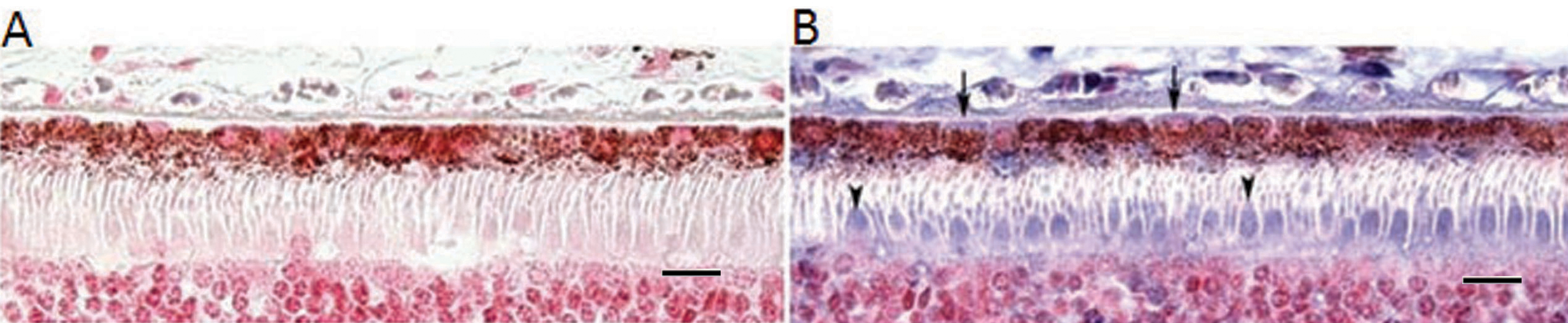
 Figure 10 of Hjelmeland, Mol Vis 2010; 16:1108-1121.