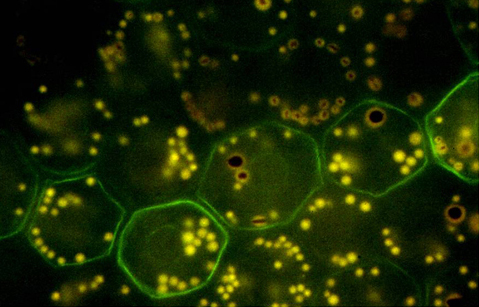
Figure 1. Bovine retinal pigment epithelium (RPE) immunolabeling for vimentin. Whole mount of a bovine retinal pigment epithelium monolayer
immunolabeled for the intermediate filament protein vimentin to illustrate a mosaic pattern of protein expression. Vimentin
has a circumferential distribution in the peripheral cytoplasm (green) within a row-like subset of retinal pigment epithelium
cells. The tissue shown here is from the tapetal region of the cow eye, which has relatively few melanosomes (brown granules),
lipofuscin (yellow granules), and combined melanolipofuscin granules [
1].
 Figure 1 of
Hjelmeland, Mol Vis 2010; 16:1108-1121.
Figure 1 of
Hjelmeland, Mol Vis 2010; 16:1108-1121.