Figure 8. Light microscopy (A, B) and transmission microscopy (C-F) of the rabbits’ trabecular meshwork. Light micrographs of all groups showed the intact geometry of the meshwork. The trabecular
meshwork (box) and Schlemm’s canal (arrow) was identified and isolated for TEM. A TEM of all groups showed a normal trabecular
meshwork and Schlemm’s canal. The trabecular beams were characterized by a predominance of transversely sectioned normal collagen
fibers in the central nucleus, widely separated by electron-lucent spaces. Elastin-like plaques varying in number and size
were also observed in all three groups. These were randomly distributed in the central nucleus of the beams and under the
basement membrane. Elongated, attenuated endothelial cells covered the outside of each beam. The inner wall of the Schlemm’s
canal revealed giant vacuoles within the endothelial cells. There was no evidence of inflammatory cells present in the trabecular
meshwork. Magnification: A, B; 100×; C, D; 2,200×; bar=5 µm; E, F; 2,800×; bar=5 µm.
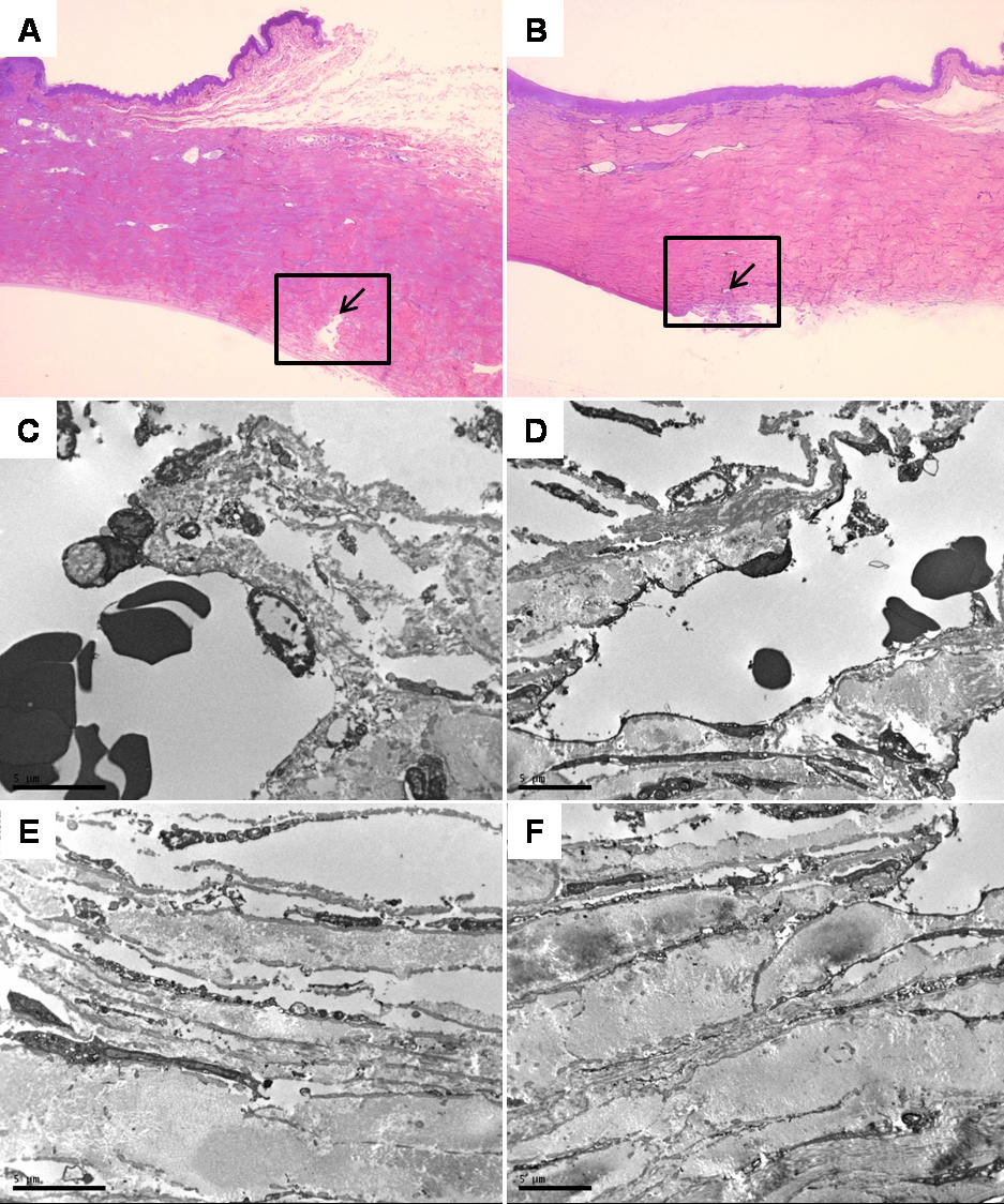
 Figure 8 of
Chew, Mol Vis 2010; 16:1087-1097.
Figure 8 of
Chew, Mol Vis 2010; 16:1087-1097.