Figure 7. Transmission electron micrographs of a representative rabbit cornea (A, B) after the fibrin glue had dissolved, showing an endothelial cell (E) adherent to Descemet’s membrane (D), with a relatively
regular cell surface and well-preserved intracellular organelles. The nucleus (N), rough endoplasmic reticulum, and mitochondria
were seen in the cytoplasm. Of note, there was no mitotic response of the endothelial cells and no evidence of endothelial
cell proliferation. Magnification: A, 7,100×; bar=1µm; B, 18,000×; bar=0.5µm.
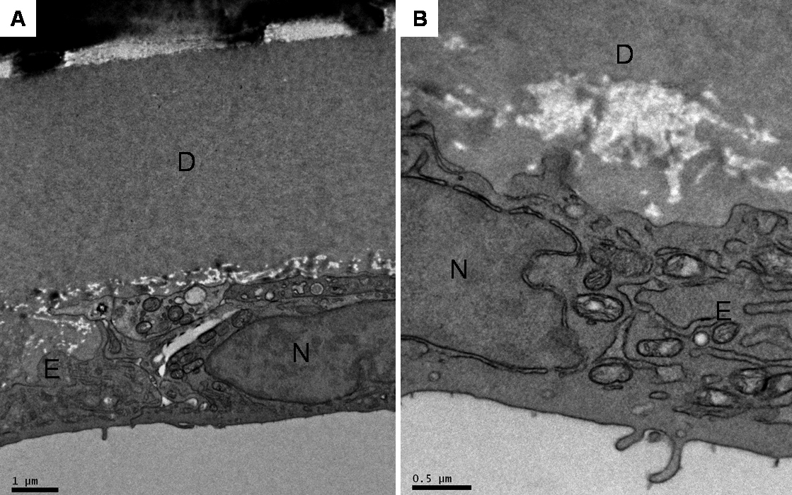
 Figure 7 of
Chew, Mol Vis 2010; 16:1087-1097.
Figure 7 of
Chew, Mol Vis 2010; 16:1087-1097.