Figure 6. Scanning electron micrographs of rabbit corneal endothelium from the three different groups (A-D). All endothelial surfaces showed a regular hexagonal cellular mosaic and well-defined cell borders, with occasional pleomorphic
cells. D: One corneal endothelium had residual TISSEEL glue attached to it (box), showing distortion to the surrounding endothelial
cells. Magnification 500×; bar=50 µm.
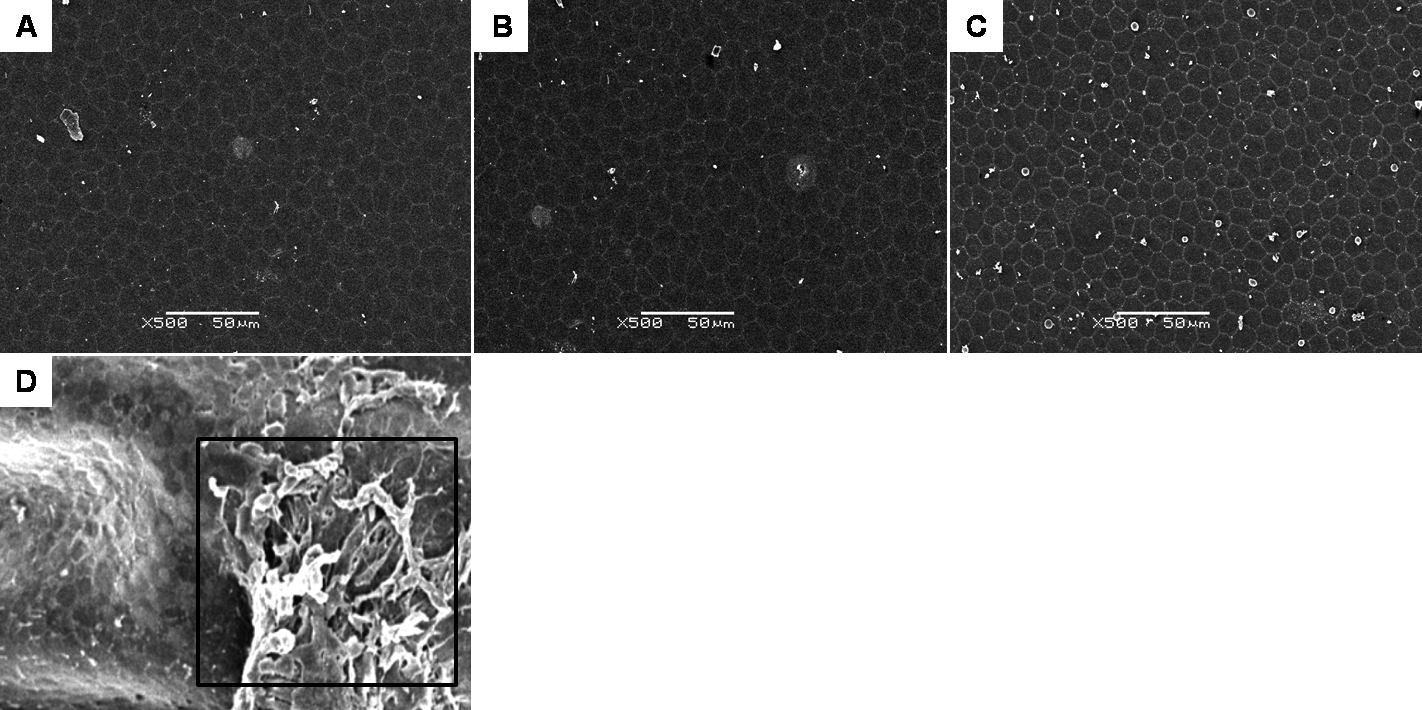
 Figure 6 of
Chew, Mol Vis 2010; 16:1087-1097.
Figure 6 of
Chew, Mol Vis 2010; 16:1087-1097.