Figure 3 of
Ji, Mol Vis 2010; 16:1068-1075.
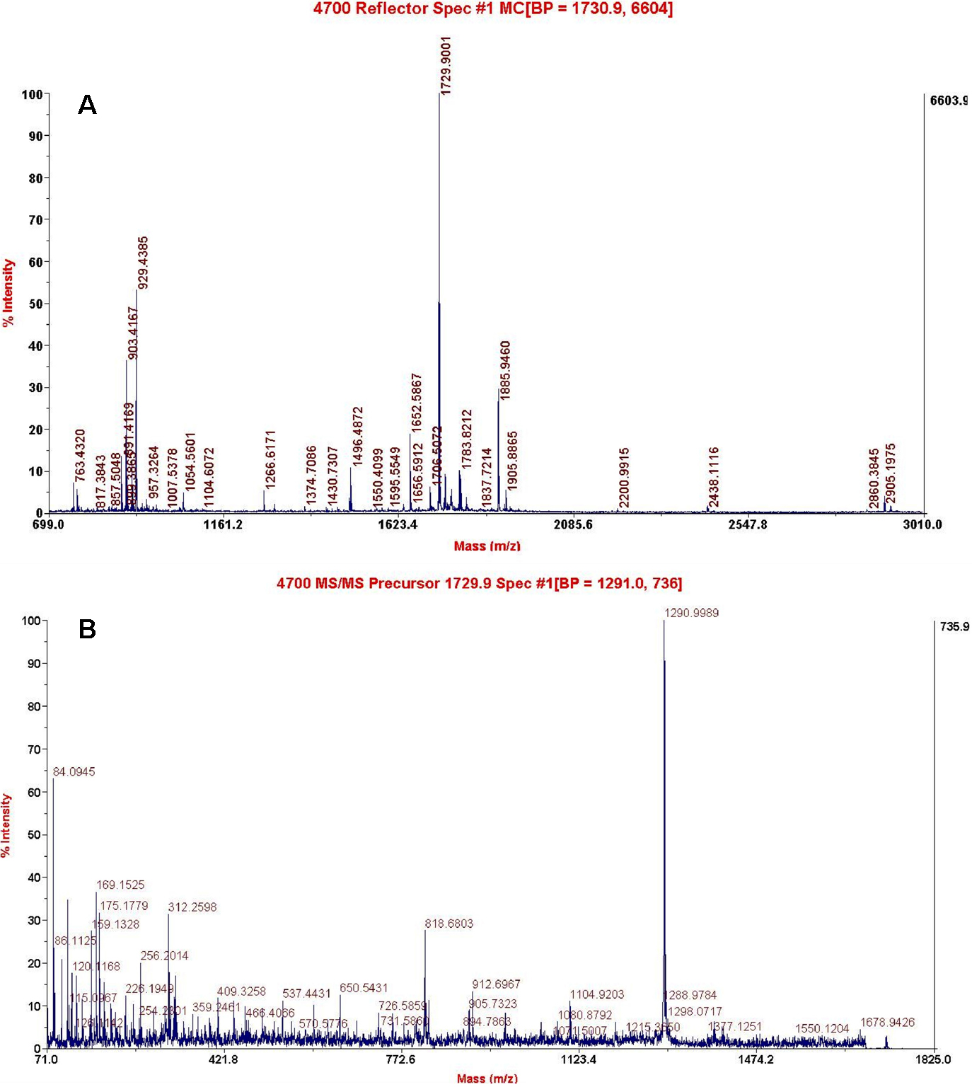
Figure 3.
Identification of mouse γS-crystallin by mass spectrometry.
A
: MALDI-MS spectrum of the γS-crystallin spot digested with trypsin.
B
: Tandem MS spectrum of the ion with m/z 1729.9 from a tryptic peptide.
 Figure 3 of
Ji, Mol Vis 2010; 16:1068-1075.
Figure 3 of
Ji, Mol Vis 2010; 16:1068-1075.