Figure 2. 2-DE gel showing the differential proteins of lenses. 190 μg of samples were loaded. Pre-cast gels of 12.5 % were used for
the second dimension, and were stained with colloid Coomassie brilliant blue (CBB) R-250. A: Six differential crystallins were identified from mutant mice. Red indicates absence (γS-crystallin) or reduction (γF-crystallin).
Blue indicates increment (βA1-, βB1-, βB2-, and αB-crystallin). B: Sixteen crystallins were identified in normal mice, including αA~αB-, βA1~βA4-, βB1~βB3-, γA~γF-, and γS-crystallin.
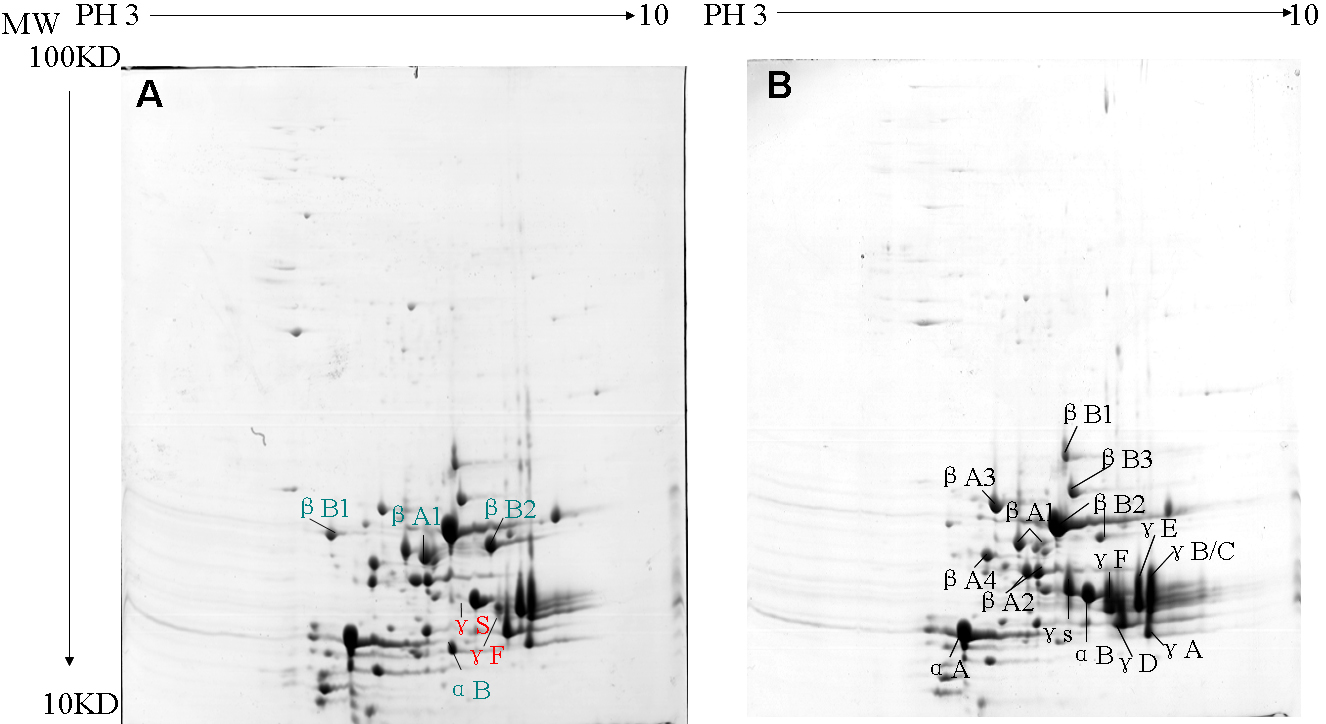
 Figure 2 of
Ji, Mol Vis 2010; 16:1068-1075.
Figure 2 of
Ji, Mol Vis 2010; 16:1068-1075.