Figure 5. Double labeling of a retina after light-exposed magnetic resonance imaging experiment. Numerous green fluorescent protein
(GFP)-positive cells (arrows, A) were immunolabelled for calbindin (B), a marker of horizontal cells. Merge image is shown in C. These data do not suggest that there were substantial numbers of infected remodeled cones (which would not be stained for
calbindin). Abbreviations are as follows: INL represents inner nuclear layer; IPL represents inner plexiform layer; GCL represents
ganglion cell layer. The scale bar is equal to 25 μm.
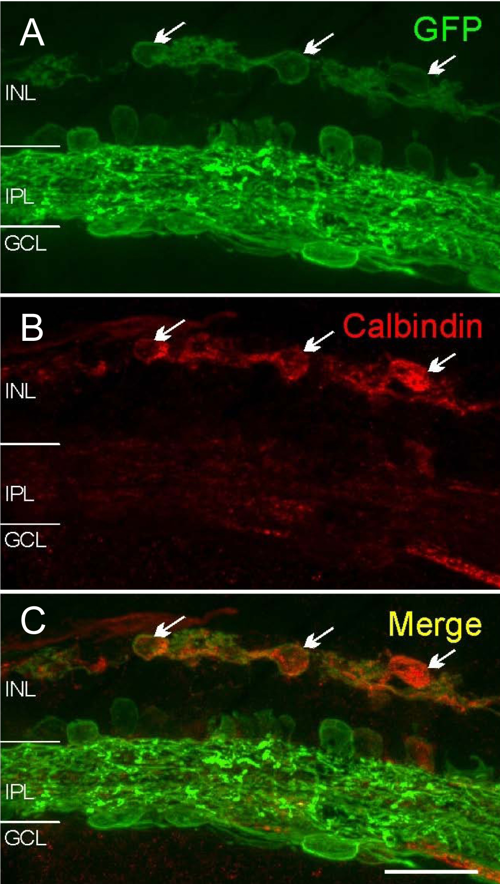
 Figure 5 of
Ivanova, Mol Vis 2010; 16:1059-1067.
Figure 5 of
Ivanova, Mol Vis 2010; 16:1059-1067.