Figure 1. Whole-mount retinas injected with channelopsin-2 viral constructs. A: A representative whole-mount retina with channelopsin-2 (Chop2-GFP) expression is shown at low resolution. A white rectangle
frames the area around the optic nerve head that was analyzed in the manganese-enhanced magnetic resonance imaging (MEMRI)
study; this area is shown magnified in E. B-H: Expression of Chop2-GFP was studied in montaged and magnified images of the retinas of dark-exposed mice (B-D) and light-exposed mice (E-H). Scale bars are 2 mm in A and 0.5 mm in B-H. Abbreviations are as follows: S represents superior, I represents inferior, N represents nasal, T represents temporal.
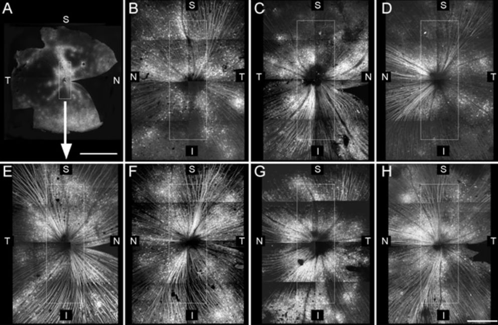
 Figure 1 of
Ivanova, Mol Vis 2010; 16:1059-1067.
Figure 1 of
Ivanova, Mol Vis 2010; 16:1059-1067.