Figure 1. Representative immunostaining results for E-cadherin associated protein expression. A: The first antibody replaced with IgG was used as the negative control. B: The representative positive β-catenin immunostaining in conjunctival tissue was used as the positive control. C: E-cadherin protein expression detected in the membrane (400×). D: β-Catenin protein expression detected in the membrane (400×). In addition, aberrant localization of β-catenin was also detected
in the cytoplasm/nuclei (E) and nucleus (F) (400×).
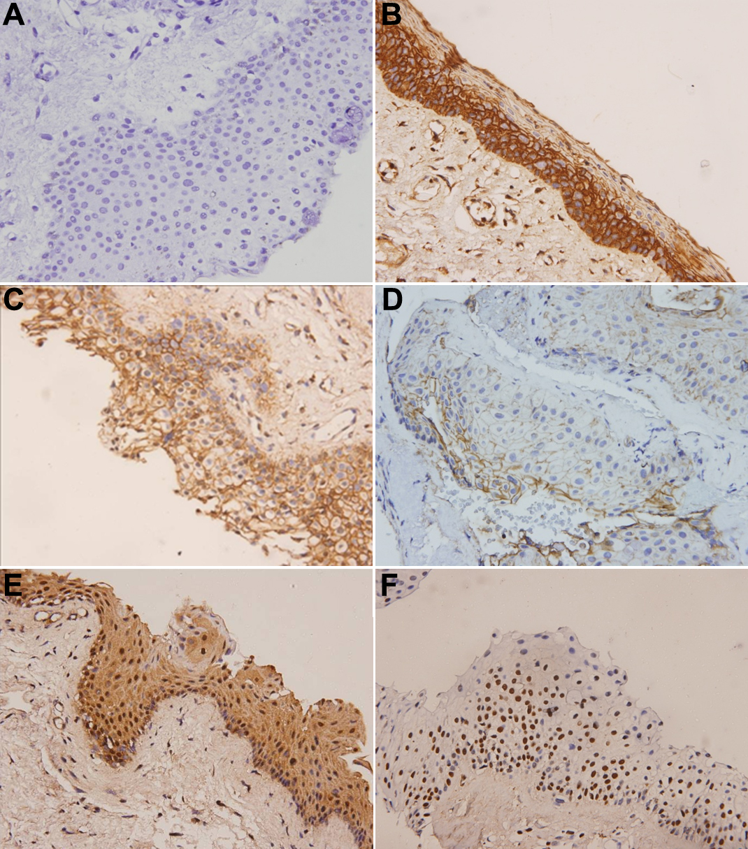
 Figure 1 of
Young, Mol Vis 2010; 16:1047-1053.
Figure 1 of
Young, Mol Vis 2010; 16:1047-1053.