Figure 5. T-cell markers were detected in
autosomal dominant neovascular inflammatory vitreoretinopathy (ADNIV)
eyes. A: Lymph node tissue is a positive control for the T-cell
marker, cluster of differentiation-3 (CD3), which was shown by the
brown color reaction. B-C: In ADNIV eyes, the
supraciliary space and vitreous showed CD3 positive cells. D-E:
In
control (non-ADNIV) phthisical eyes, CD3 positive cells were absent.
F: Lymph node tissue is a positive control for the T-cell
marker, cluster of differentiation-4 (CD4), which was shown by the
brown color reaction. G-H: In ADNIV eyes, the
supraciliary space and vitreous both showed CD4 positive cells. I-J:
In
control (non-ADNIV) phthisical eyes, CD4 positive cells were absent.
K: Lymph node tissue is a positive control for the T-cell
marker, cluster of differentiation-8 (CD8), which was shown by the
brown color reaction. L: In ADNIV eyes, the supraciliary space
showed CD8 positive cells. M: In the ADNIV vitreous, CD8
positive cells were absent. N-O: In control (non-ADNIV)
phthisical eyes, CD8 positive cells were absent. The scale bar
represents 50 μm.
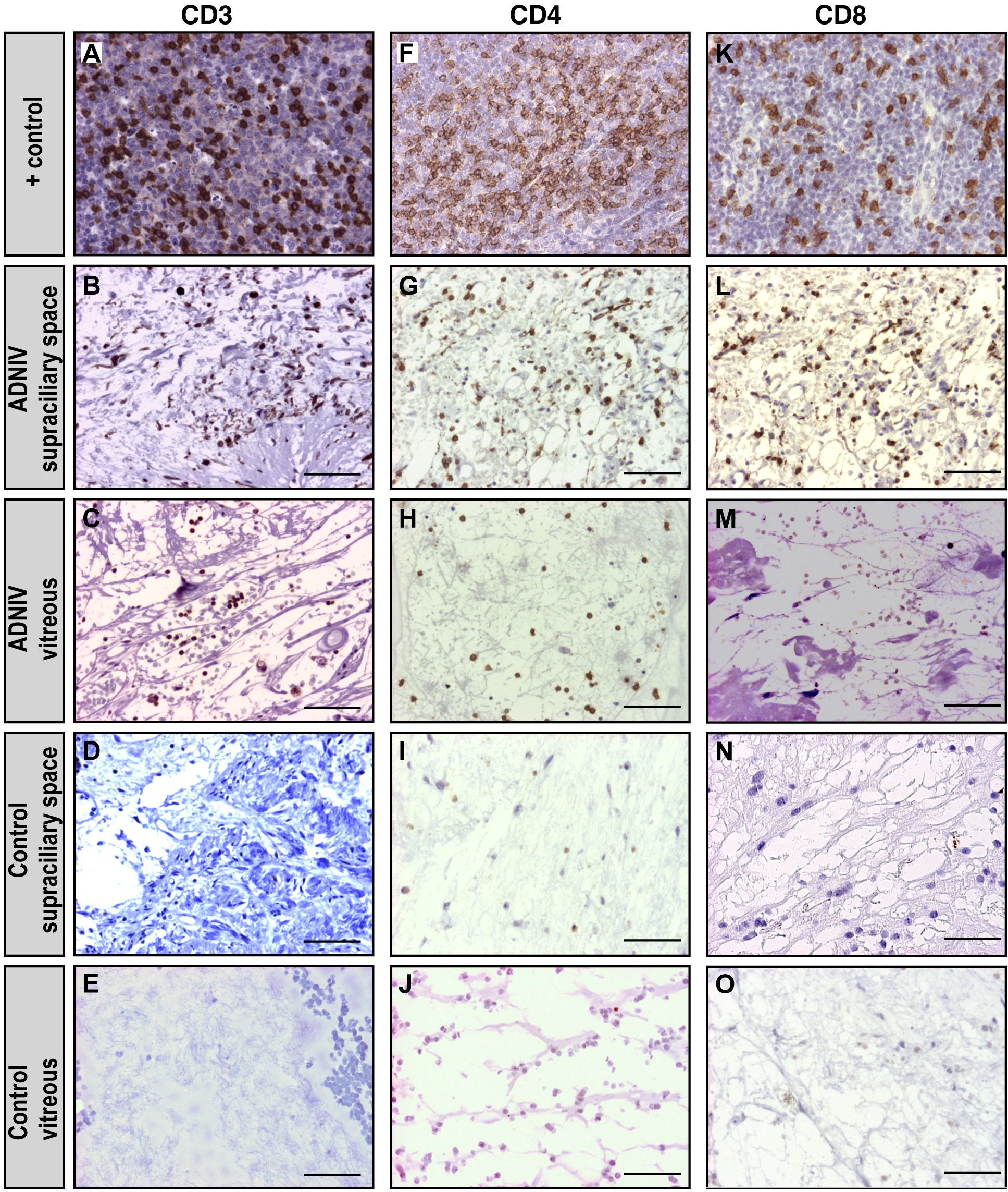
 Figure 5 of Mahajan, Mol Vis 2010; 16:1034-1040.