Figure 2. Masson’s trichrome stain of
autosomal dominant neovascular inflammatory vitreoretinopathy (ADNIV)
eye demonstrates fibrovascular proliferation and features of phthisis. A:
There
was massive fibrovascular connective tissue within the pupil,
overlying the iris, and causing detachment of the ciliary body. B:
A
supraciliary effusion was present outside the detached ciliary body. C:
The
sclera was thickened. D: There were a few lens remnants
present. E: A fibrovascular stalk extended from the optic nerve
to the peripheral retina. F: The retina was detached, atrophic,
and osseous metaplasia was present.
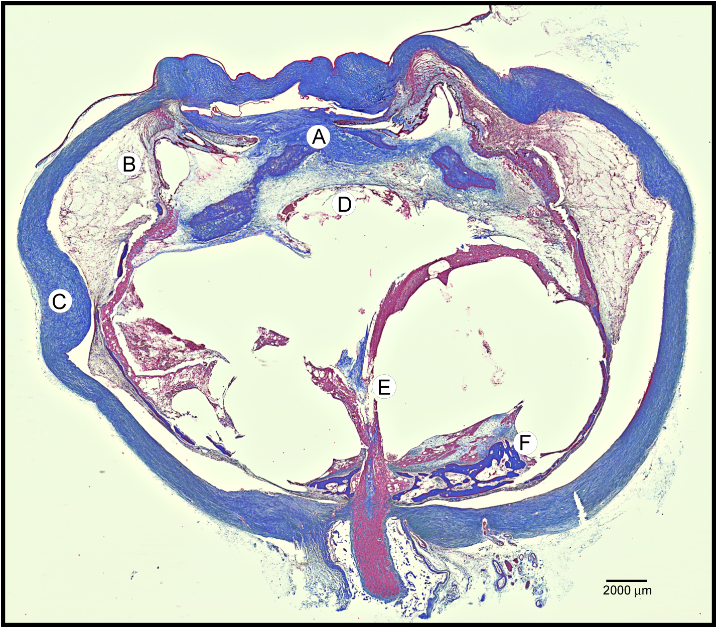
 Figure 2 of Mahajan, Mol Vis 2010; 16:1034-1040.