Figure 1. End-stage autosomal dominant neovascular inflammatory vitreoretinopathy. A: This phthisical right eye shows cloudy corneas, iris neovascularization, and pupillary fibrous membrane with aphakia. B: The B-scan demonstrates a foreshortened disorganized eye with total retinal detachment and calcification. C: The fellow prephthisical eye showed a neovascular stalk on the optic disc. D: Electroretinogram reveals extinguished photopic and scotopic responses.
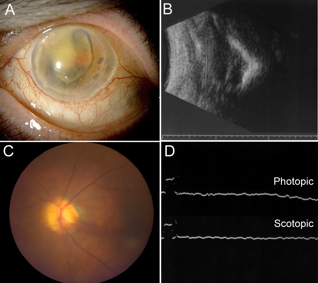
 Figure 1 of
Mahajan, Mol Vis 2010; 16:1034-1040.
Figure 1 of
Mahajan, Mol Vis 2010; 16:1034-1040.