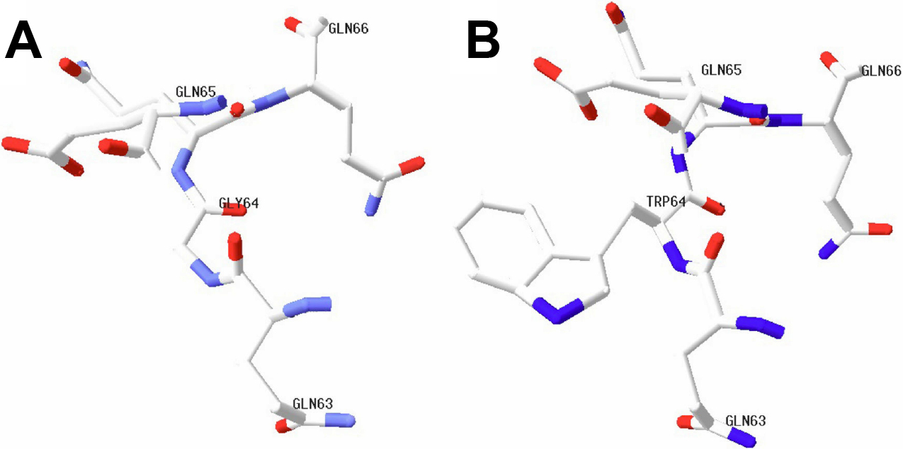
Figure 5. Portion of the crystallin, beta A4 protein (CRYBA4) modeled structure in the vicinity of residue 64. The portion modeled structures
of the CRYBA4 in the vicinity of residue 64 were built using Swiss-PdbViewer 4.0.1 [
39-
41]).
A: The normal modeled structure of CRYBA4 is displayed.
B: The mutant modeled structure of CRYBA4 is shown. Glycine (Gly) is replaced by tryptophan (Trp) at codon 64. Modeled structures
are shown by element type, using a default standard CPK (a popular color convention for distinguishing atoms of different
chemical elements) scheme: n=blue, O=red, C=white.
 Figure 5 of
Zhou, Mol Vis 2010; 16:1019-1024.
Figure 5 of
Zhou, Mol Vis 2010; 16:1019-1024.