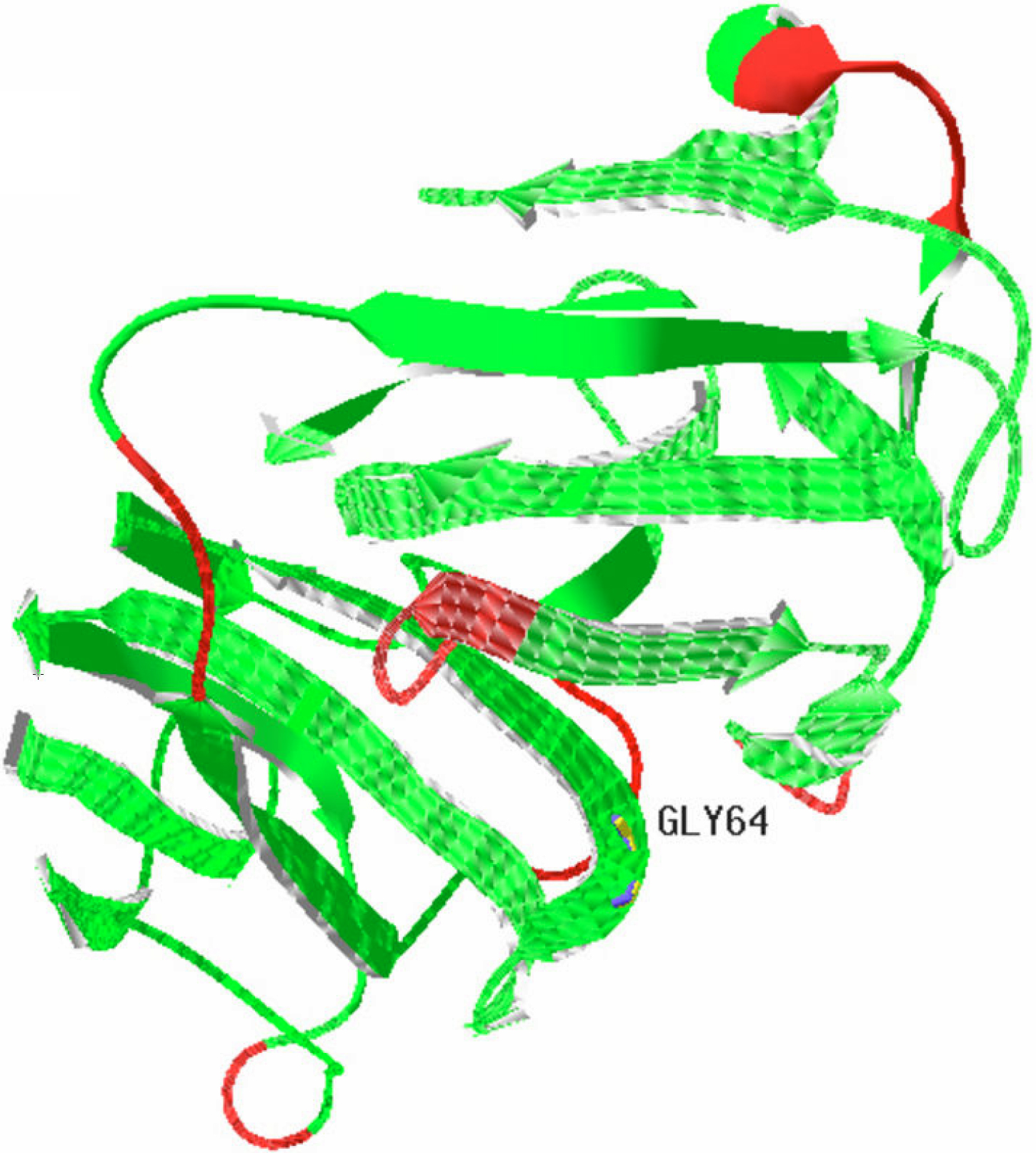
Figure 4. The modeled structure of
crystallin, beta A4 protein (CRYBA4). The modeled structure of CRYBA4
was built using special software (Swiss-PdbViewer 4.0.1 [
39-
41]). The mutation
described in this study leads to the replacement of glycine by
tryptophan at codon 64 (Gly64).The Gly64, located in the corner of the
modeled structure, may form such secondary structures as β-sheet or
β-turn.
 Figure 4 of Zhou, Mol Vis 2010; 16:1019-1024.