Figure 5. Immunoreactivities of matrix
metalloproteinase-2 (MMP-2) and MMP-9. A-D:
Immunohistochemical labeling of MMP-2 (A) and MMP-9 (C)
in normal control retinas. Retinal photocoagulation resulted in
increased MMP-2 (B) and MMP-9 (D) expression in the
retinas. Immunoreactivities of cluster differentiation 44 (CD44) and
CD44s. E–F: Immunohistochemical labeling of CD44 (E)
and
CD44s (G) in normal control retinas. Immunohistochemical
labeling of CD44 in laser injured retinas (F). Retinal
photocoagulation resulted in increased expression of CD44s (H),
the degradation product of CD44. Nuclei are counter stained with
To-Pro3. Scale bar represents 75 µm.
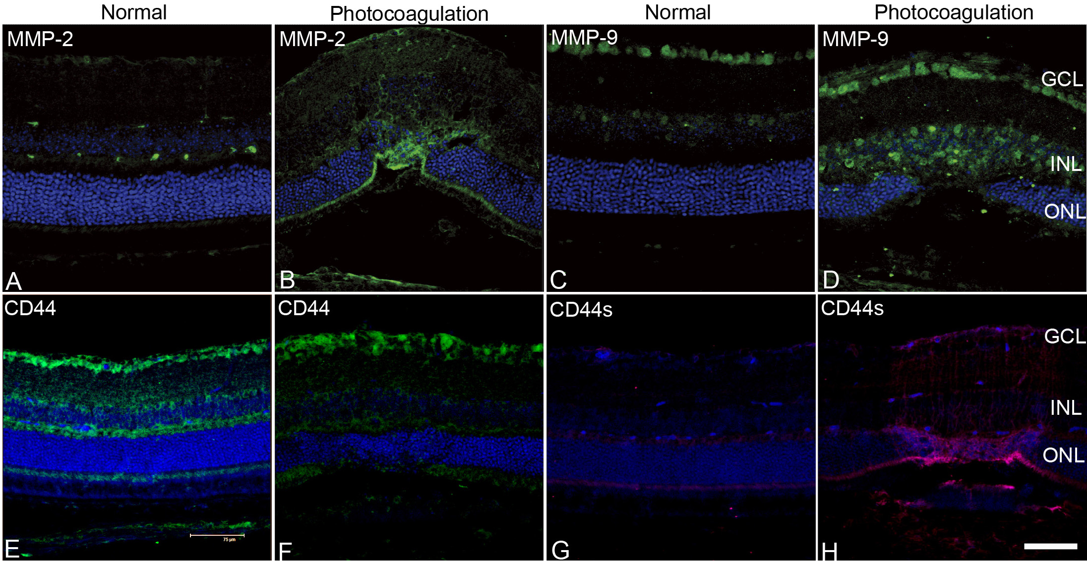
 Figure 5 of Jiang, Mol Vis 2010; 16:983-990.