Figure 3. Newly developed outer segments
expressed rhodopsin. Following transplantation, donor RPCs integrated
into the outer nuclear layer (ONL) and developed outer segments
co-labeled with green fluorescent protein (GFP; A), outer
segment marker (rhodopsin; B). Panel C is a merged
image of the two panels to the left. Scale bar represents 4 μm.
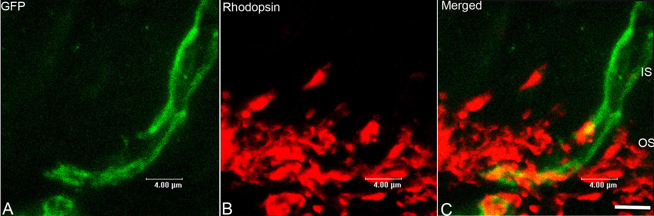
 Figure 3 of Jiang, Mol Vis 2010; 16:983-990.