Figure 4. The 2M6 antigen is not expressed
by neurons of the chick retina. Hatchling chick retina was fixed,
embedded in paraffin, sectioned, and stained with antibodies specific
for 2M6 (red) and calbindin (blue) and with peanut agglutinin (green).
Panel A shows the full extent of the retina and retinal
pigmented epithelium with 2M6 and peanut agglutinin signals merged with
a 4',6-diamidino-2-phenylindole (DAPI; amber) labeling of cell nuclei.
Panels B and C show higher magnification views at the
outer and inner extremes of the neural retina, respectively.
Abbreviations are as follows: PC represents the photoreceptor cell
layer; INL represents the inner nuclear layer; IPL represents the inner
plexiform layer; GC/NF represents the ganglion cell/nerve fiber layer.
The magnification bar for A is 41.6 μm; the magnification bar
for B is 16 μm; the magnification bar for C is 5.39 μm.
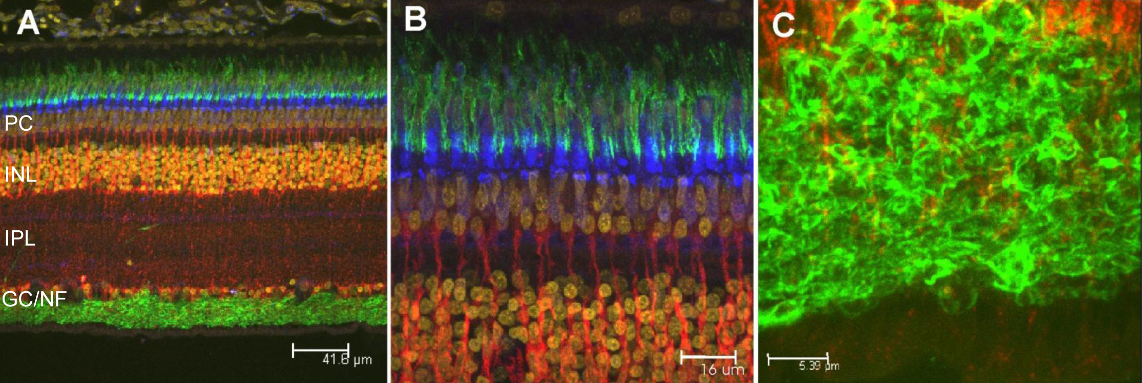
 Figure 4 of Ochrietor, Mol Vis 2010; 16:961-969.