Figure 3. The 2M6 antigen is an
intracellular protein expressed by Müller glial cells of the chick
retina. Hatchling chick retina was fixed, embedded in paraffin,
sectioned, and stained with antibodies specific for 2M6 (A, red)
and 5A11/Basigin (B, green), and neurofilaments (C,
blue). The overlay image (D) includes
4',6-diamidino-2-phenylindole (DAPI) staining of DNA (amber). Note that
2M6, which is expressed by Müller cells, is most heavily distributed
between the ganglion cell layer and the outer limiting membrane. In
contrast, 5A11/Basigin, also expressed by Müller cells, extends beyond
the ganglion cells into the nerve fiber layer to the Müller cell
endfeet. In panels E through H, higher magnification
images compare and contrast the intracellular Müller cell membrane
staining of 2M6 (red) with the Müller cell plasma membrane staining of
5A11/Basigin (green). In panel E, the Müller cell perikarial
region of the inner plexiform layer is shown. In panel F, the
Müller cell bodies are shown. Müller cell processes at the outer
plexiform/photoreceptor cell layer are shown in panel G,
whereas Müller cell processes at the ganglion cell/nerve fiber layer
are shown in panel H. Abbreviations are as follows: PC
represents the photoreceptor cell layer; OPL represents the outer
plexiform layer; INL represents the inner nuclear layer; IPL represents
the inner plexiform layer; GC/NF represents the ganglion cell/nerve
fiber layer. The magnification bars for A–D are 23.6 μm;
the magnification bar for E is 9.3 μm; the magnification bars
for F–H are 3.4 μm.
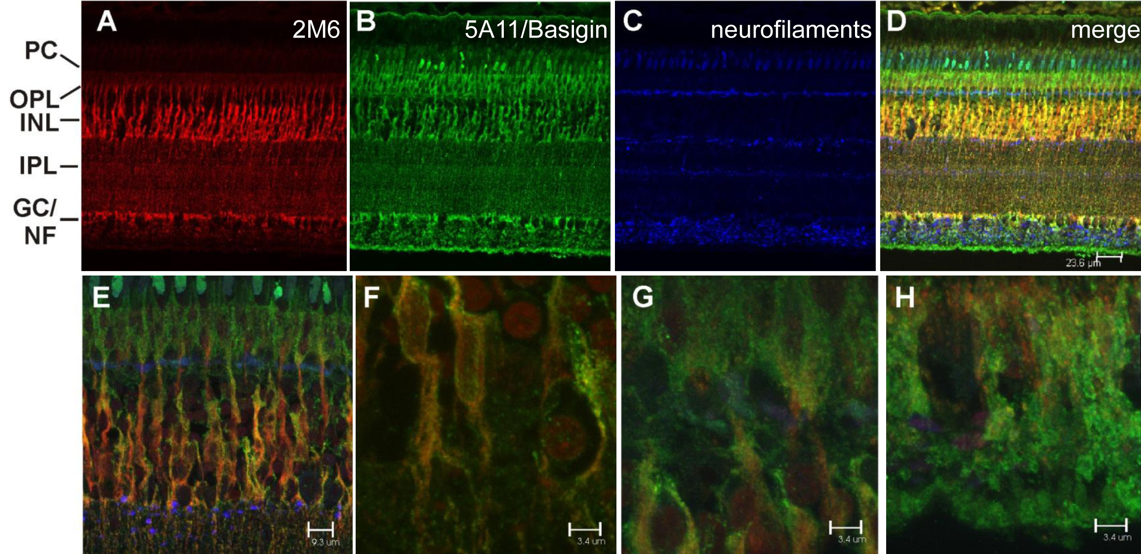
 Figure 3 of Ochrietor, Mol Vis 2010; 16:961-969.