Figure 3 of
Clausen, Mol Vis 2010; 16:954-960.
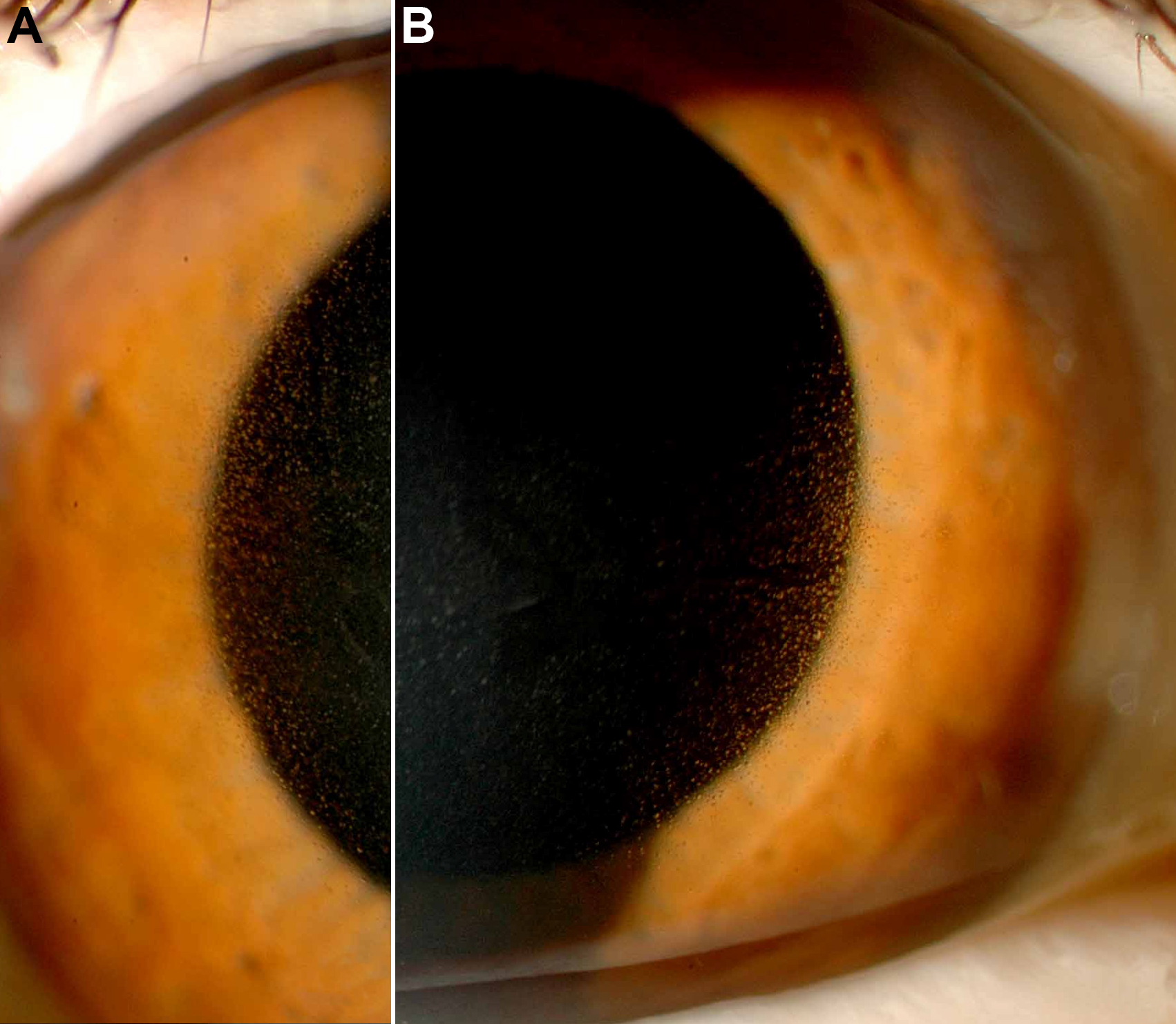
Figure 3.
Slit lamp photography of patient I/2. The corneal epithelium showed bilateral myriads of fine cysts predominantly in the center of the cornea (
A
=right eye,
B
=left eye).
 Figure 3 of Clausen, Mol Vis 2010; 16:954-960.