Figure 1. Isolation and characterization
of side population (SP) cells from the mouse lens epithelium.
Fluorescence-activated cell sorting (FACS) analysis of mouse lens
epithelial cells stained with Hoechst 33342 alone (A) or in the
presence of verapamil (B). The P1 region shows the gated region
identified SP cells. The P2 region is designated as non-SP cells. SP
cells (C) and non-SP cells (D) were observed by phase
contrast microscopy. The scale bar represents 10 µm. (E) Forward
scatter characteristics (FSC) of SP cells (solid area) and non-SP cells
(open area) were analyzed by FACS.
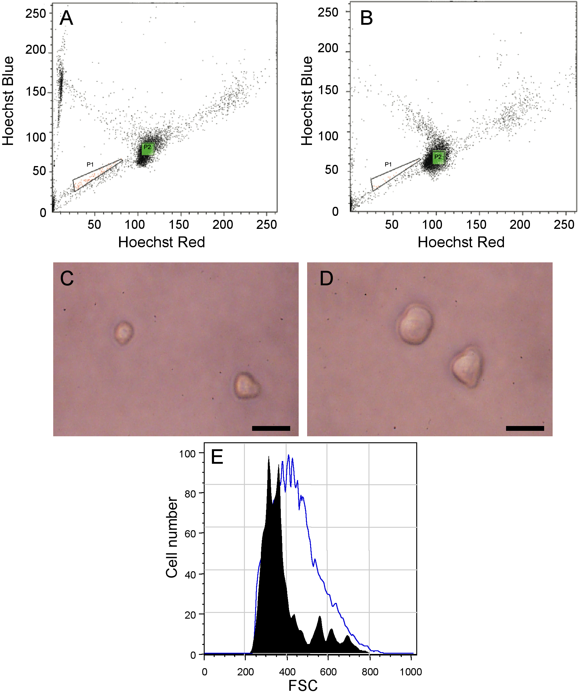
 Figure 1 of Oka, Mol Vis 2010; 16:945-953.