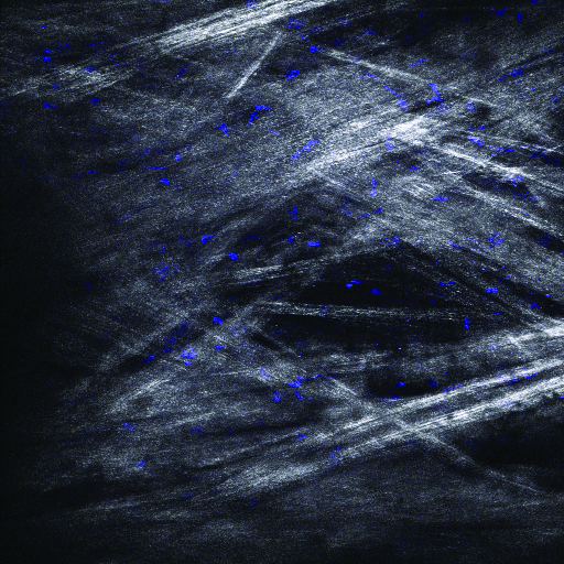
 Figure 8 of Ammar, Mol Vis 2010; 16:935-944.
  Figure 8 of Ammar, Mol Vis 2010; 16:935-944.
Note that the slide bar at the bottom of the quicktime movie can be used to manually control the flow of the movie, or double click on the image to automatically run the animation. If you are unable to view the movies, a representative frame from each movie is included below.
| This animation requires Quicktime 6 or
later. Quicktime is available as a free download. |
