Figure 6. Cellular specificity of the
pFIN-IRBP156-CHER-RK-GFP-WPRE dual-promoter vector.
pFIN-IRBP156-CHER-RK-GFP-WPRE (1.5×10⁹ vector genomes/µl)
lentivirus was injected into the developing neural tubes of E2 chicken
embryos in ovo. The retinas of the injected embryos were harvested on
E19–20, examined as whole mounts using native fluorescent, and
sectioned (10 µm). A-C: A representative section showing
the extent of photoreceptor infection. The merged image indicates that
nearly all cells are co-expressing the two fluorescent reporter
proteins, CHER and GFP. D-F: Close up of transduced
photoreceptor layer in region containing INL cells expressing the viral
transgene (arrow in D). All sections were counterstained with
DAPI. All scale bars shown equal 50 µm. Abbreviations: ONL represents
outer nuclear layer; INL represents inner nuclear layer; IPL represents
inner plexiform layer.
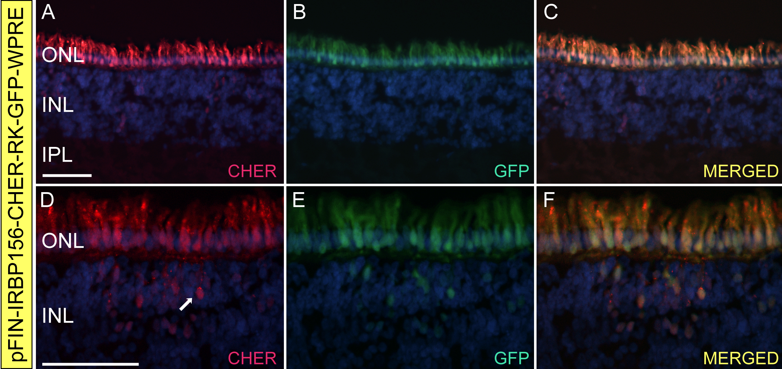
 Figure 6 of Semple-Rowland, Mol Vis 2010; 16:916-934.