Figure 1. Cellular activities of
interphotoreceptor retinoid binding protein promoter (IRBP156), Xenopus
opsin promoter (XOPS), neural retina leucine zipper (NRLL), and
rhodopsin kinase (RK) promoters in chicken retina. Lentiviral vectors
were injected into the ventricles of chicken embryos (embryonic day 2
–E2) in ovo. The viruses injected were as follows: A-B
pFIN-IRBP156-tdTOM; C-D pFIN-XOPS-tdTOM; E-F
pFIN-NRLL-tdTOM; G-H pFIN-RK-GFP-WPRE. The retinas of the
injected embryos were harvested on E19–20 and the cells expressing the
fluorescent reporter proteins were identified using native fluorescent
and immunofluorescent microscopy. In selecting the representative
images shown in this and subsequent figures, our goal was to document
all of the cell types and the variability in expression levels of the
reporter proteins observed in transduced cell populations. The sections
shown in B, D, F, and H were
immunostained with a rod transducin polyclonal antibody that was
visualized using either goat anti-rabbit Alexa Fluor 488 (B, D,
F) or 594 (H) secondary antibody. Arrows indicate inner
retinal cells (A) or rod photoreceptors (B, H).
All sections were counterstained with DAPI. All scale bars shown equal
50 µm. Abbreviations: ONL represents outer nuclear layer; INL
represents inner nuclear layer.
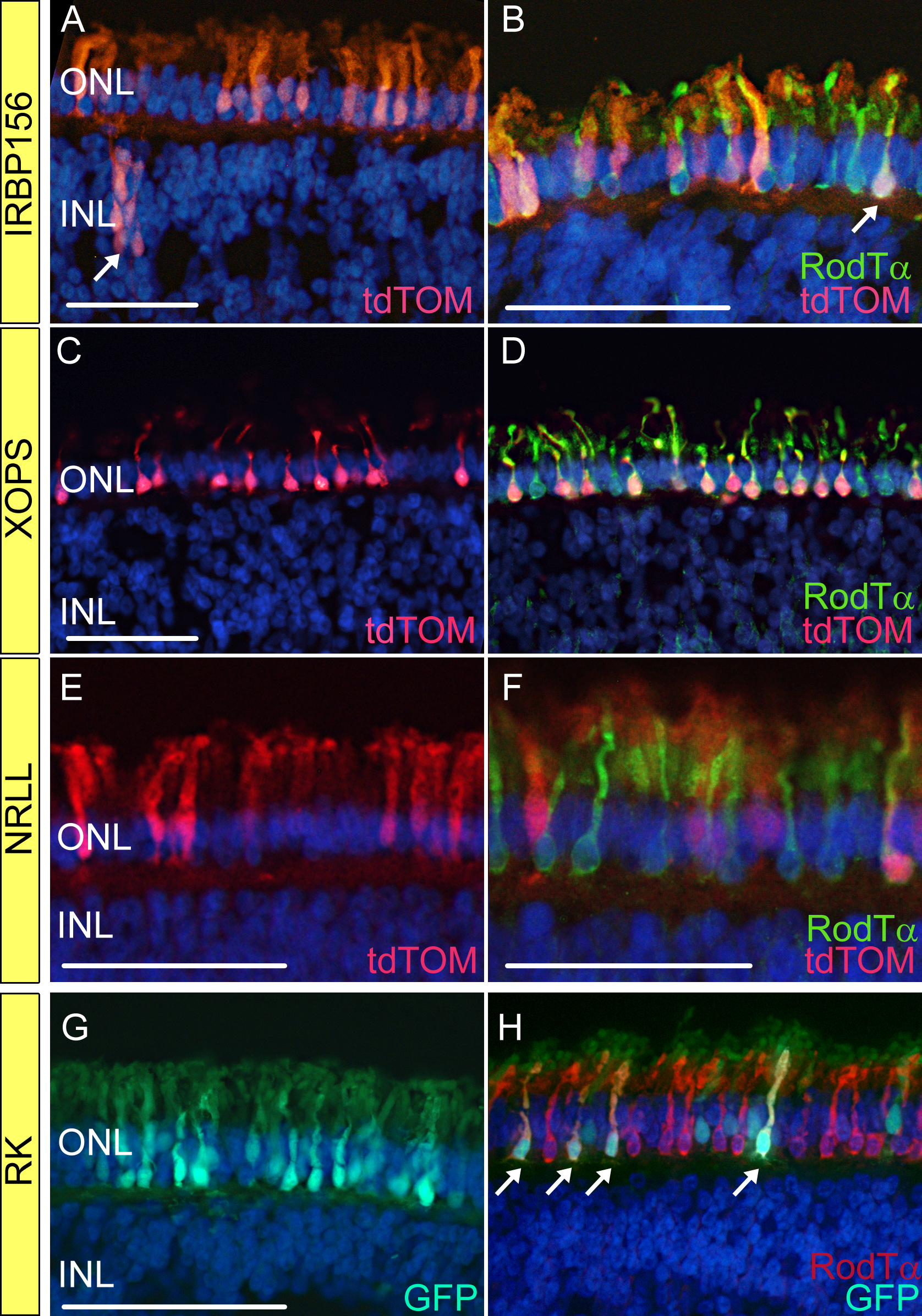
 Figure 1 of Semple-Rowland, Mol Vis 2010; 16:916-934.