Figure 5. Representative fluorescence
microscopic images of actively cycling HCEC from a 73-year-old donor.
HCEC were electroporated with control or p21+p16 siRNA and then seeded
onto multi-well chamber slides. On Days 1, 5, and 11 after
electroporation, cells were immunostained for Ki67 (green) to view
actively cycling cells and with DAPI (blue) to visualize all nuclei.
Arrowheads indicate Ki67-positive dividing cells. Original
magnification was 20×.
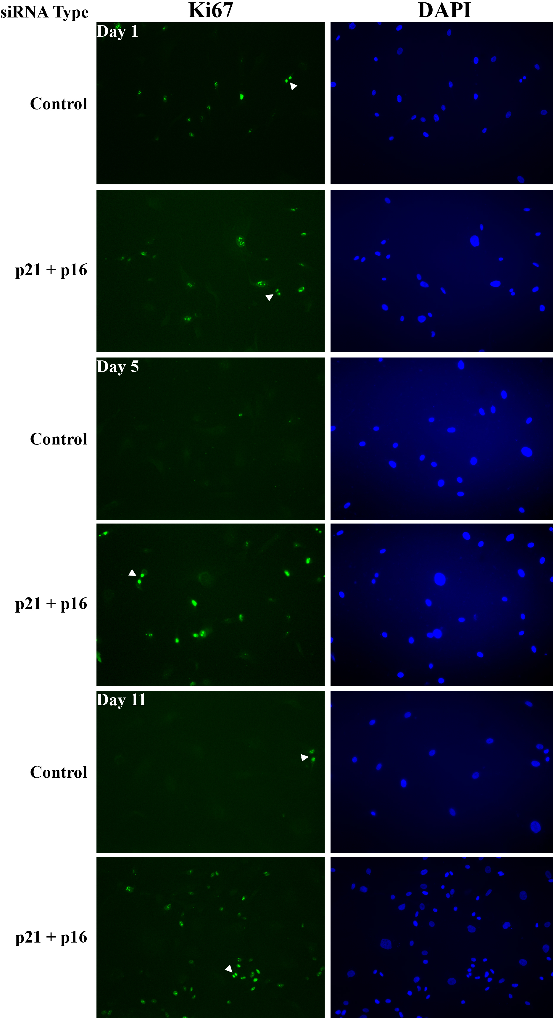
 Figure 5 of Joyce, Mol Vis 2010; 16:897-906.