Figure 3. Lysozyme and albumin
distribution through lotrafilcon B. CLSM (confocal laser scanning
microscopy) scans were analyzed to locate the fluorescent-conjugated
protein on the front surface, within the bulk region, and on the back
surface of lotrafilcon B after one and 14 days of incubation. Lenses
were either rinsed in phosphate buffered saline (PBS), soaked overnight
in hydrogen peroxide (H2O2) or soaked overnight
in a multipurpose solution (MPS) with (MPS-RUB) or without (MPS-NO-RUB)
manual lens rubbing. A, B: These panels show the
results for HEL (hen egg lysozyme), C, D: These panels
show the results for BSA (bovine serum albumin).
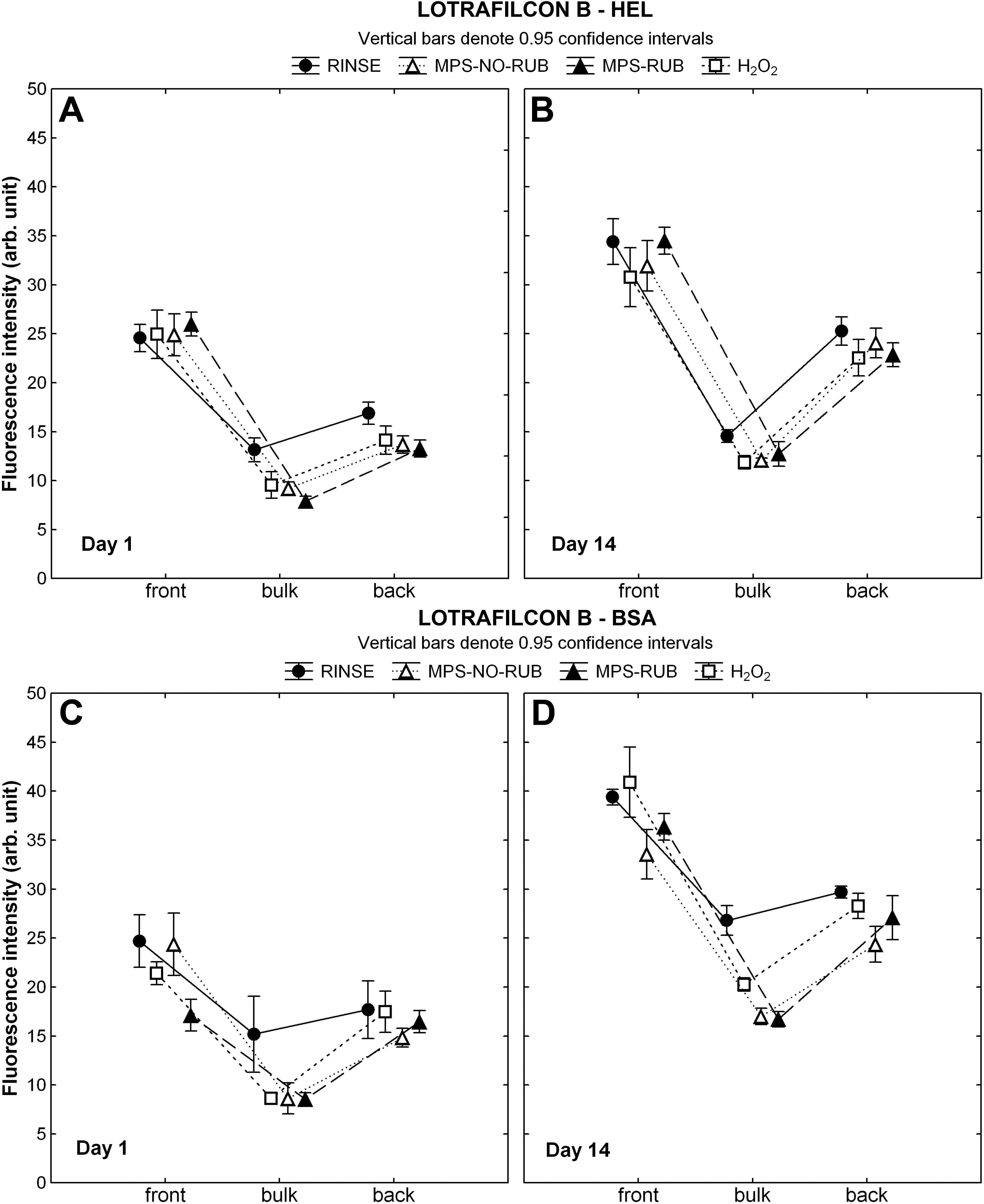
 Figure 3 of Luensmann, Mol Vis 2010; 16:79-92.