Figure 1. Schematic diagram for
experimental procedures. Contact lenses were incubated in lysozyme
(HEL) and albumin (BSA) solution, followed by overnight
soaking in different care regimens and the two methods to locate and
quantify the protein on the lens. In the figure, 125I indicates iodine125;
BSA
indicates bovine serum albumin; CL indicates contact lens; CLSM
designates confocal laser scanning microscope; Exp indicates
Experiment; H2O2 indicates hydrogen peroxide; HEL
designates hen egg lysozyme; LY designates lucifer yellow vinyl
sulfone; and MPS indicates multipurpose solution.
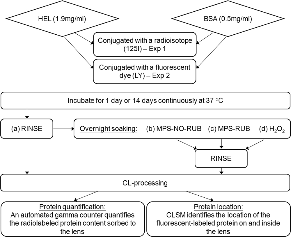
 Figure 1 of Luensmann, Mol Vis 2010; 16:79-92.