Figure 2. Histological examination of the
paraffin sections stained with hematoxylin and eosin. A: The
diffusely swelled collagen fibers were twisted or frayed (arrows) and
mixed with normal fibers in the sclera (Subject III15; magnification,
400X). B: The alignment of collagen fibers was disrupted, and
there were a lot of coil-like amorphism materials (arrows) in the
sclera of an atrophied eye globe (Subject III10; size, 20 mmx18 mmx15
mm; magnification, 100X).
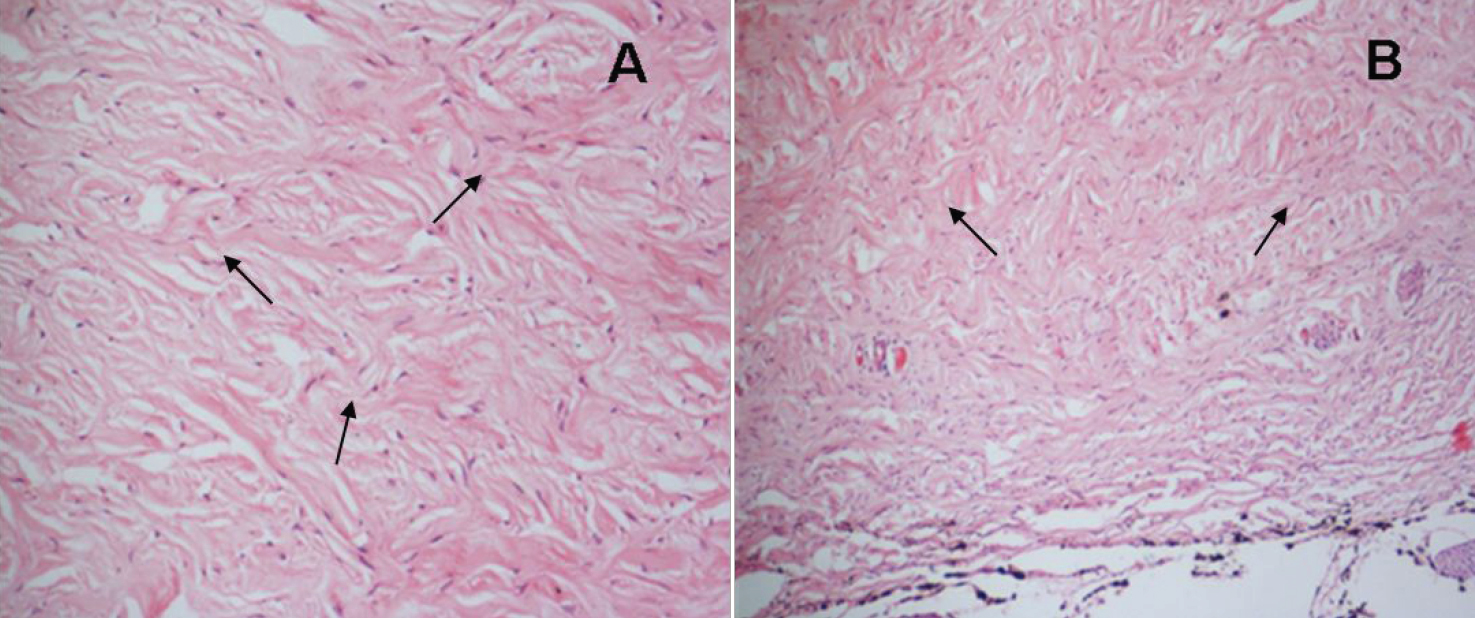
 Figure 2 of Yu, Mol Vis 2009; 15:949-954.