Figure 6. Immunohistochemical staining for NFκB and IRF3 in HCECs stimulated with poly(I:C) and cultured with or without of DEX or CsA
for 24 h. NFκB p65 staining without poly(I:C, A), with poly(I:C, B), with DEX 10-5M and poly(I:C, C), and with CsA 10-5M and poly(I:C, D). IRF3 staining without poly(I:C, E), with poly(I:C, F), with DEX 10-5M and poly(I:C, G), and with CsA 10-5M and poly(I:C, H). Scale bar, 100 µm. Activated NFκB p65 and IRF-3 were clearly detected in the nuclei of most of cultured HCECs 3 h after
stimulation by poly(I:C, B and F). In the presence of DEX, NFκB p65 and IRF-3 were detected in the nuclei of some HCECs but only in the cytosol of other HCECs
(C, G). In the presence of CsA, NFκB p65 staining was detected in more HCEC nuclei after exposure to CsA than to DEX (D), while
IRF3 was detected only in the nuclei of cultured HCECs (H).
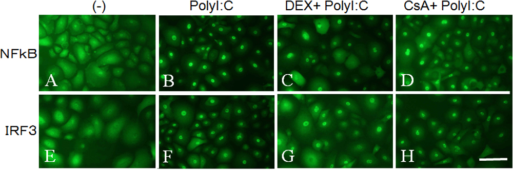
 Figure 6 of
Hara, Mol Vis 2009; 15:937-948.
Figure 6 of
Hara, Mol Vis 2009; 15:937-948.