Figure 4. Sections of human retina
immunocytochemically labeled for GH and dying cells. A: A
section from a 74 year-old male labeled with DAPI (blue) for nuclei,
growth hormone (GH) antiserum (red; asterisk); and TUNEL (green) for
apoptotic nuclei. B: Another section from the same individual,
labeled in the same way as A. C: A section from an 81
year-old male, labeled in the same way as A. D: A
section from a 69 year-old male, labeled in the same way as A.
With respect to cells in the ganglion cell layer (GCL), in the merged
images, non-apoptotic and GH-positive nuclei appear red (asterisks) and
correspond to nuclei numbered: A 5, B 2, C 1, D
2, and D 4. Apoptotic and GH-negative nuclei A 2, A
4, B 1, B 4, B 5, C 2, D 1, and
D 3, appear blue-green. Non-apoptotic and GH-negative nuclei A
1, A 3, and B 3 appear blue. Abbreviations: inner
nuclear layer (INL); outer nuclear layer (ONL). Scale bar equals 20 μm.
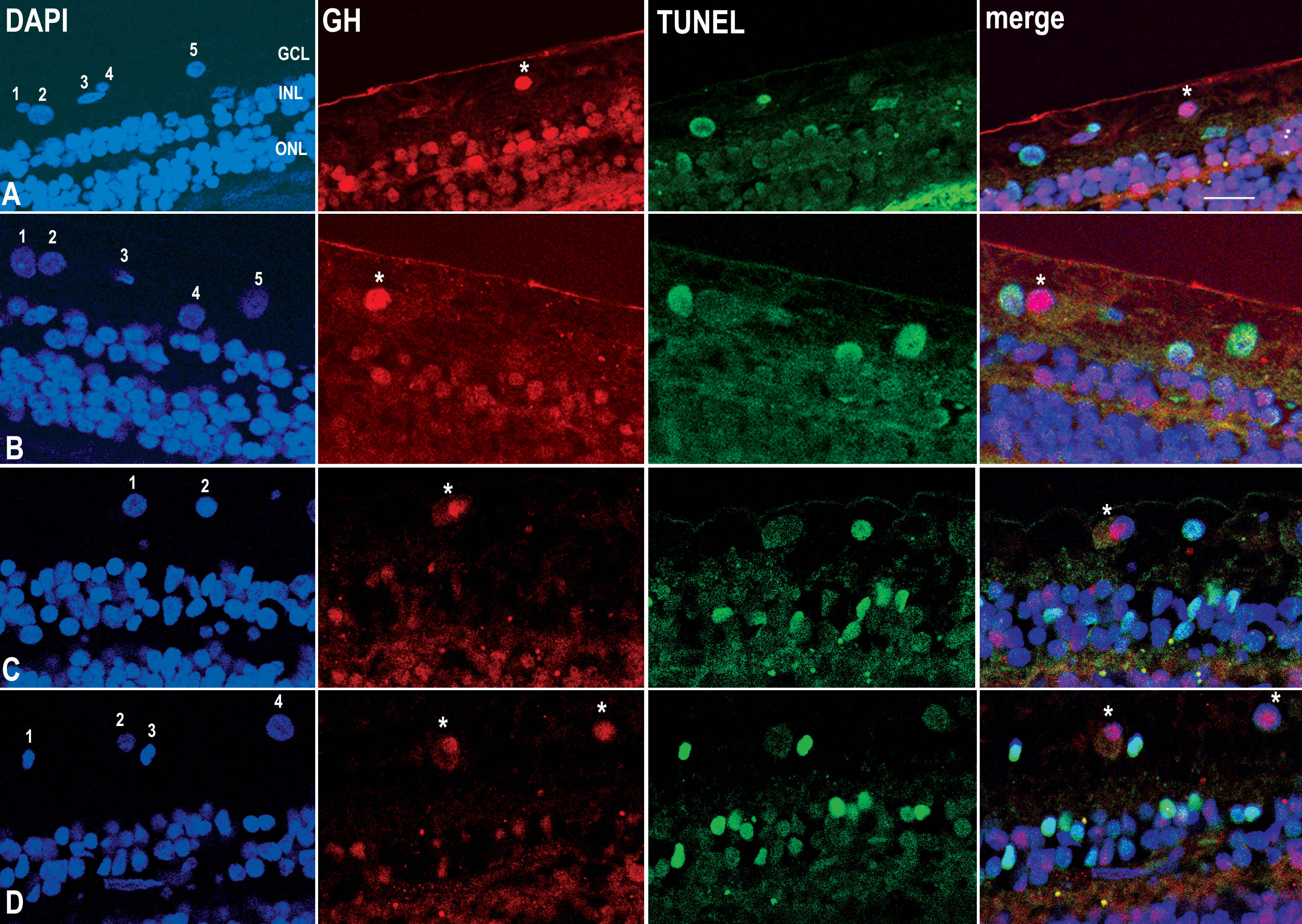
 Figure 4 of Sanders, Mol Vis 2009; 15:920-926.