Figure 3. A human retina section double-labeled for γ-synuclein and GH. A: γ-synuclein antibodies (red) label a retinal ganglion cell (RGC) in the ganglion cell layer (GCL). B: growth hormone (GH) antiserum (green) labels a nucleus in the GCL. C: The merged image of panels A and B shows that GH localizes to the same cell as γ-synuclein, an RGC marker, (asterisks) indicating that this GH-containing cell
is an RGC. DAPI labeling (blue) shows the position of all nuclei in the section. Abbreviations: ganglion cell layer (GCL);
inner nuclear layer (INL). Scale bar equals 20 μm.
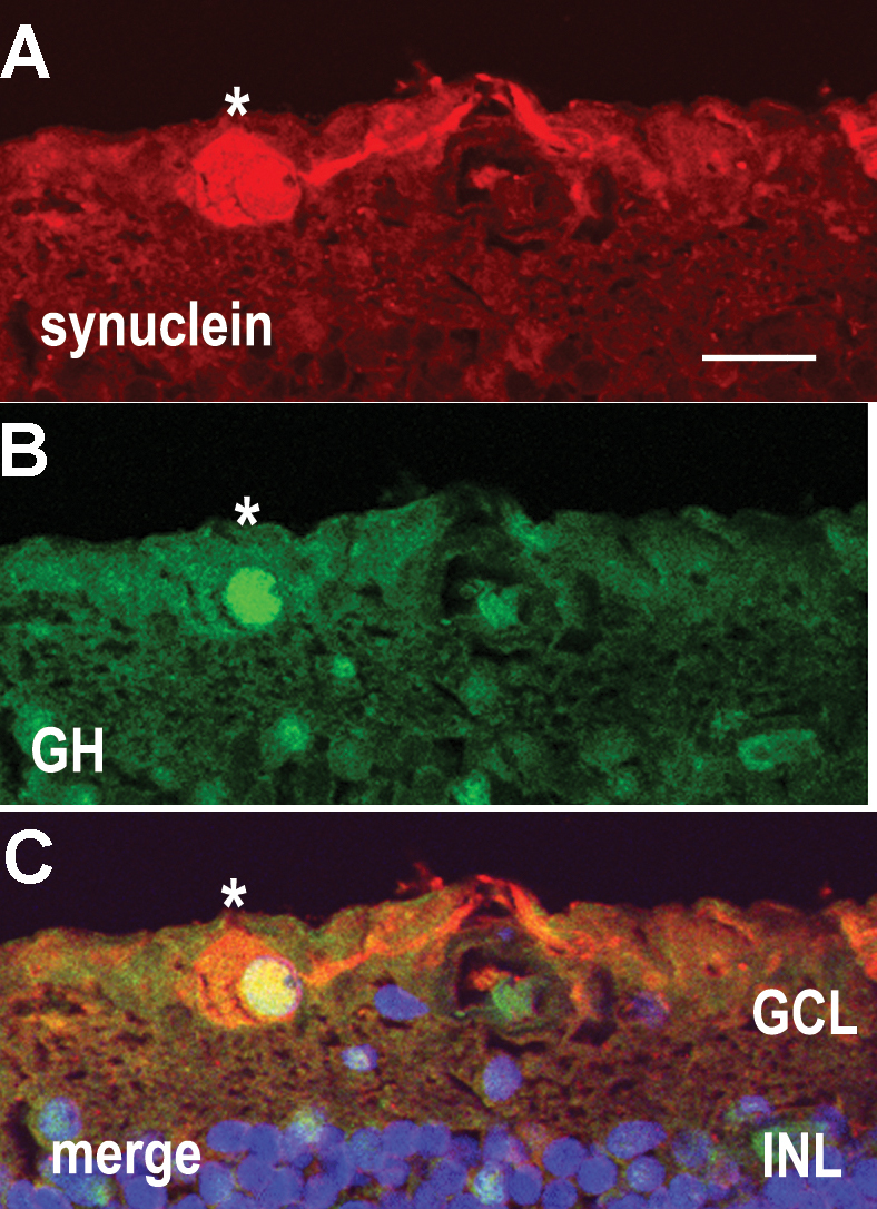
 Figure 3 of
Sanders, Mol Vis 2009; 15:920-926.
Figure 3 of
Sanders, Mol Vis 2009; 15:920-926.