Figure 2. Human retina sections showing an immunocytochemical control and γ-synuclein labeling. A: This image shows immunocytochemical labeling of a section in which growth hormone (GH) antiserum was replaced by rabbit serum
and GH receptor (GHR) antibodies were replaced by mouse IgG. DAPI labeling (blue) shows the position of all nuclei in the
section. B: This is an oblique section through the retina, labeled with an antibody against γ-synuclein (green) to identify retinal ganglion
cells (RGCs) in the ganglion cell layer (GCL). The majority of cells in the GCL show cytoplasmic labeling for synuclein. DAPI
labeling (blue) shows the position of all nuclei in the section. Abbreviations: inner nuclear layer (INL); outer nuclear layer
(ONL). Scale bar equals 40 μm.
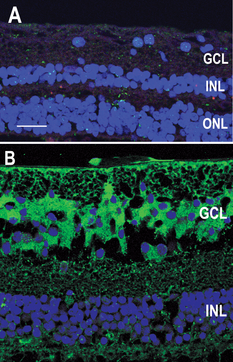
 Figure 2 of
Sanders, Mol Vis 2009; 15:920-926.
Figure 2 of
Sanders, Mol Vis 2009; 15:920-926.