Figure 2. Controls. Panel A is
negative control. Drusen (indicated by arrow in AMD) shows no binding
when the lectin was replaced by 0.05 M TBS with mM calcium chloride.
Arrowhead indicates RPE. Panel B is positive control. Normal
kidney (glomerulus indicated by dashed arrow) stains with CON A.
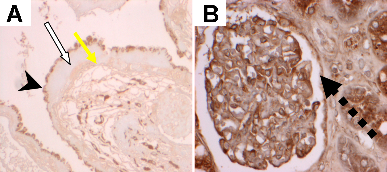
 Figure 2 of D’Souza, Mol Vis 2009; 15:906-911.