Figure 1. Comparison of the lectin-binding
pattern of drusen, RPE, Bruch’s membrane, and photoreceptors. A:
PSA binds outer segments and cell membranes of both cones (dotted
arrow) and rods (solid arrow) strongly. B: PSA binds drusen
(white arrow) and Bruch’s membrane (yellow arrow) strongly. C:
MPA binds outer segments and cell membranes of both cones (dotted
arrow) and rods (solid arrow) with moderate intensity. D: MPA
binds drusen (white arrow) and Bruch’s membrane (yellow arrow) with
moderate intensity. E: AHA without pretreatment with
neuraminidase binds outer segments and cell membranes of cones (dotted
arrow) but not rods. F: AHA without pretreatment with
neuraminidase does not bind drusen (arrow) or Bruch’s membrane. G:
After pretreatment with neuraminidase, AHA binds outer segments and
cell membranes of both cones (dotted arrow) and rods (solid, black
arrow) strongly and (H) drusen (white arrow) with moderate
intensity. Arrowhead indicates RPE. Original magnification is 600×
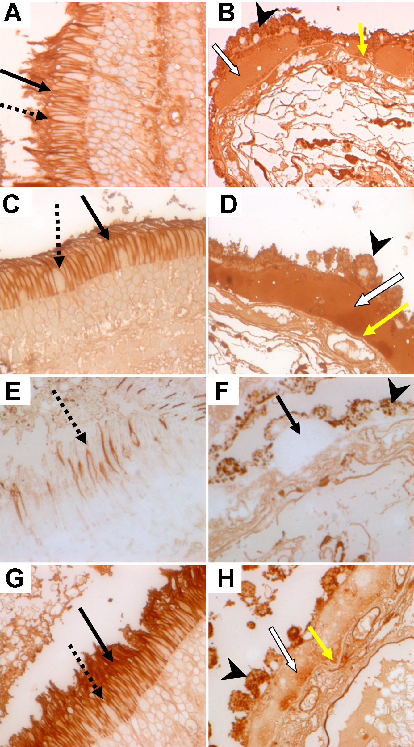
 Figure 1 of D’Souza, Mol Vis 2009; 15:906-911.