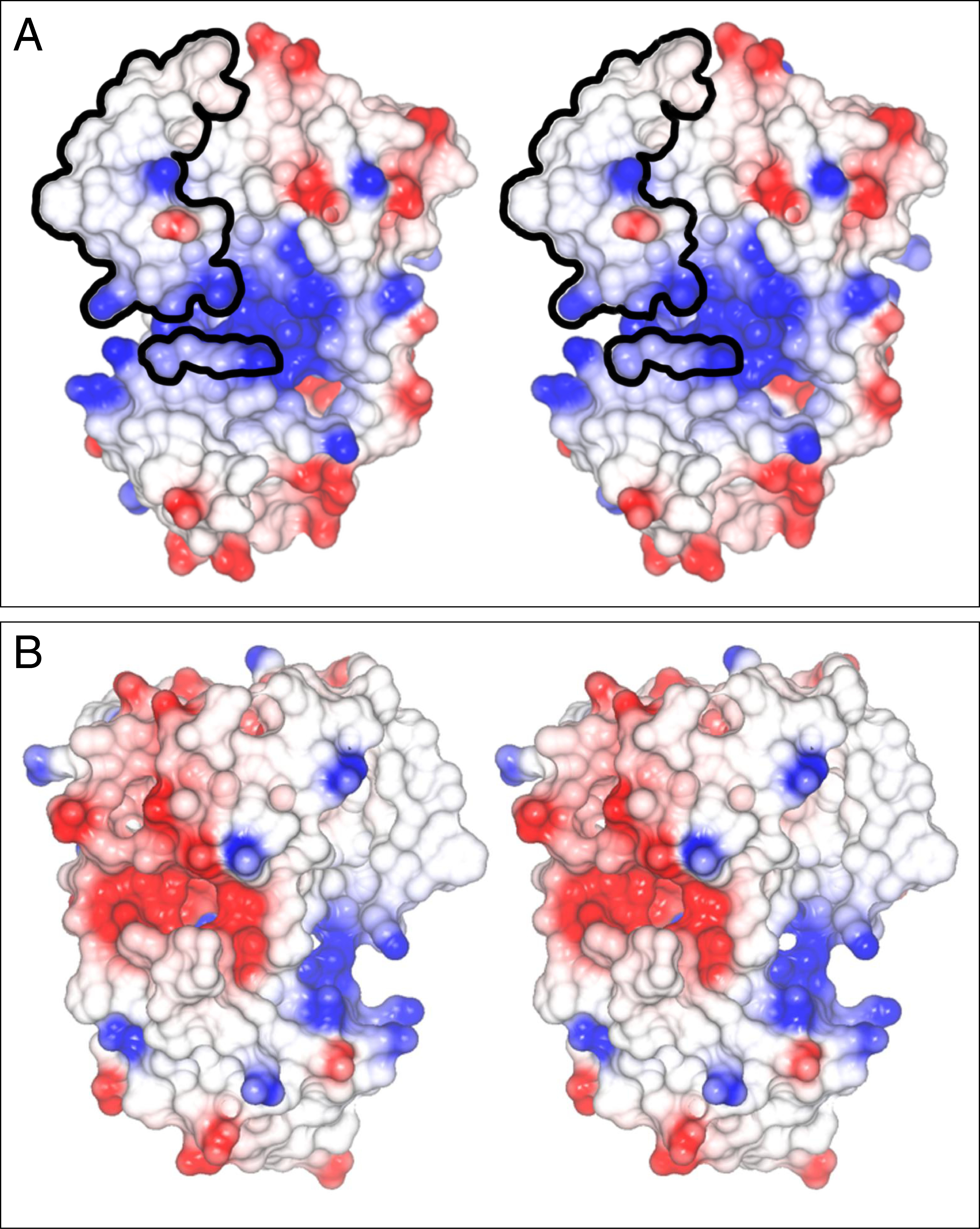
Figure 6. Electrostatic surface potential
of CRALBP. The structural model of cellular retinaldehyde-binding
protein (CRALBP; PDB entry 1XGH) is that of Liu et al. [
40]. Surface regions
in red have negative electrostatic potential and are acidic. Those in
white have neutral electrostatic potential and those in blue have
positive electrostatic potential and are basic.
A: The two
frames form a stereo pair of the basic face of CRALBP. The basic recess
(blue) between the upper and lower lobes of CRALBP is approximately 10
Å in diameter. An Arg-Ala-Arg (RAR) sequence, conserved throughout the
CRAL_TRIO protein family, is shown (small patch of circled residues).
The retinoid-binding site is covered by the lipid exchange loop (large
patch of circled residues). The basic recess may mediate interactions
of CRALBP with specific acidic lipids.
B: The two images form a
stereopair of the acidic face of the protein. Note the (negative)
acidic patch (red).
 Figure 6 of Saari, Mol Vis 2009; 15:844-854.
  Figure 6 of Saari, Mol Vis 2009; 15:844-854.