Figure 4. Release of 11-cis- and 9-cis-retinal
from CRALBP. A: Cellular retinaldehyde-binding protein
(CRALBP)·11-cis-retinal (6 μM) was incubated in the dark with 2
mM NH2OH and small unilamellar vesicles (SUVs). The total
phospholipid concentration was 90 μM, and SUVs were composed of 100
mol% phosphatidylcholine (PC) or 50 mol% PC and 50 mol% other lipids,
as indicated. After 16 h at room temperature, retinoids were extracted
and quantified by high performance liquid chromatography. The amount of
11-cis-retinal oximes (syn+anti) reflected the release of 11-cis-retinal
for reaction with NH2OH during the incubation. The amount of
11-cis-retinal released from by PC: phosphatidic acid (PA) was
approximately 75% of the total bound to CRALBP. The results shown are
from a single experiment. B: CRALBP·9-cis-retinal (6 μM)
was incubated in the dark with NH2OH and SUVs as described.
Spectra were obtained before and 3 h after addition of the SUVs. The
results are shown as the % of the maximum spectral change resulting
from incubation with 4 M urea. Error bars shown are standard deviations
from the means (n=3). The abbreviations used are: phosphatidic acid
(PA); phosphatidylcholine (PC); phosphatidylethanolamine (PE);
phosphatidylinositol (PI); phosphatidylserine (PS).
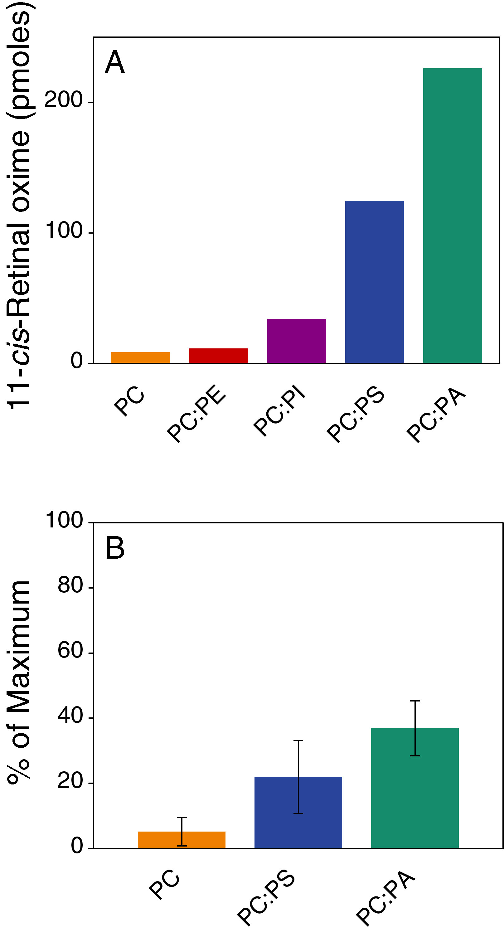
 Figure 4 of Saari, Mol Vis 2009; 15:844-854.