Figure 1. Lipid-immunoblot assay. A: Several lipids were surveyed. Lipids were dissolved in CHCl3 and applied to nitrocellulose membranes in small spots (25 nmol/spot). After the membrane was appropriately blocked and washed,
it was probed with bovine cellular retinaldehyde-binding protein (CRALBP) and anti-CRALBP as described in Methods. CRALBP
bound tightly to surfaces coated with the acidic glycerophospholipids, phosphatidic acid (PA), phosphatidylinositol-3,4,5-tris-phosphate
[PI(3,4,5)P3], phosphatidylserine (PS), phosphatidyl-4-phosphate (PI(4)P), and more weakly to surfaces coated with the other acidic lipids
phosphatidylinositol (PI), phosphatidylinositol-4,5-bisphosphate (PI(4,5)P2), and a mixture of sulfatides (Sulf). No binding was apparent with phosphatidylcholine (PC), phosphatidylethanolamine (PE),
sphingomyelin (SPH), or a mixture of gangliosides (Gang). Each of the three columns in the figure was derived from a separate
immunoblot. The size of the lipid spots was variable due to components in the lipid solutions that affected wetting of the
nitrocellulose. The results shown are representative of at least three independent analyses of each lipid. B: Active lipids were compared on the same blot. The lipids indicated below the blot were applied to nitrocellulose in small
spots (25 nmol/spot) and probed with bovine CRALBP as described for A. The order of staining intensity (PA>PS>PI>PC) was observed in at least three independent analyses. Solvent is abbreviated
Sol.
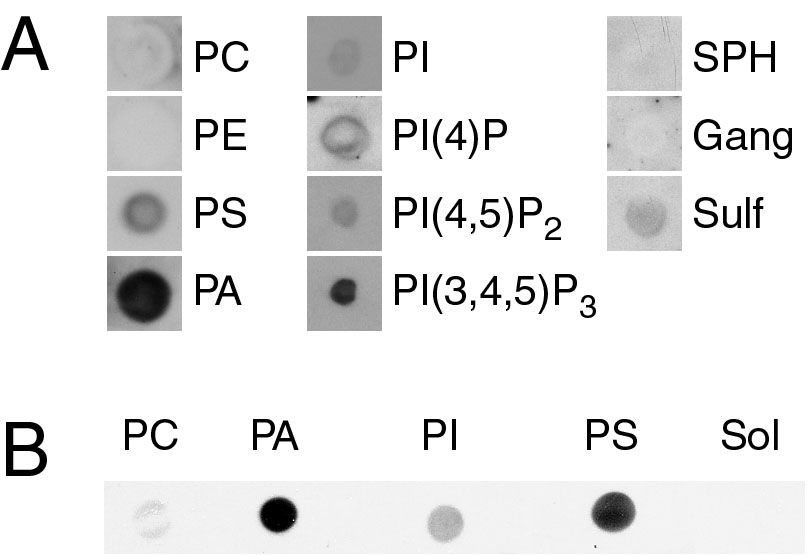
 Figure 1 of
Saari, Mol Vis 2009; 15:844-854.
Figure 1 of
Saari, Mol Vis 2009; 15:844-854.