Figure 1. Ocular findings in a Korean XLRS
patient (case 16). A: Fundus photograph of the both eyes showed
typical stellate pattern of schisis cavities in the macula. The inset
presents an image of the macula magnified twofold. B:
Fluorescein angiogram showed no definite leakage from the cystic
cavities. C: Optical coherence tomography showed the schisis in
the nerve fiber layer. D: Electroretinogram showed markedly
decreased amplitude of b-wave and relative preservation of a-wave,
which are key features of XLRS.
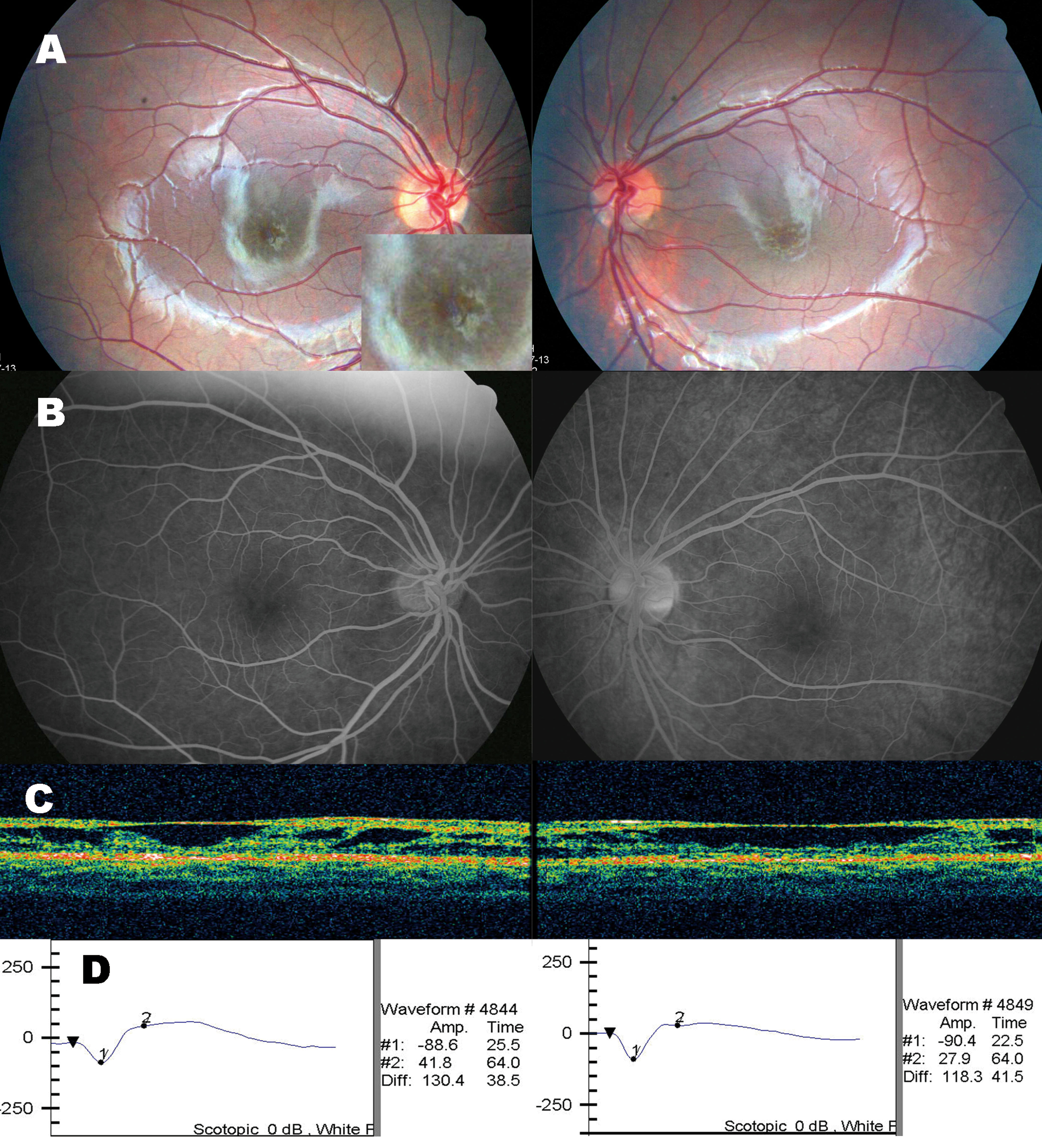
 Figure 1 of Kim, Mol Vis 2009; 15:833-843.