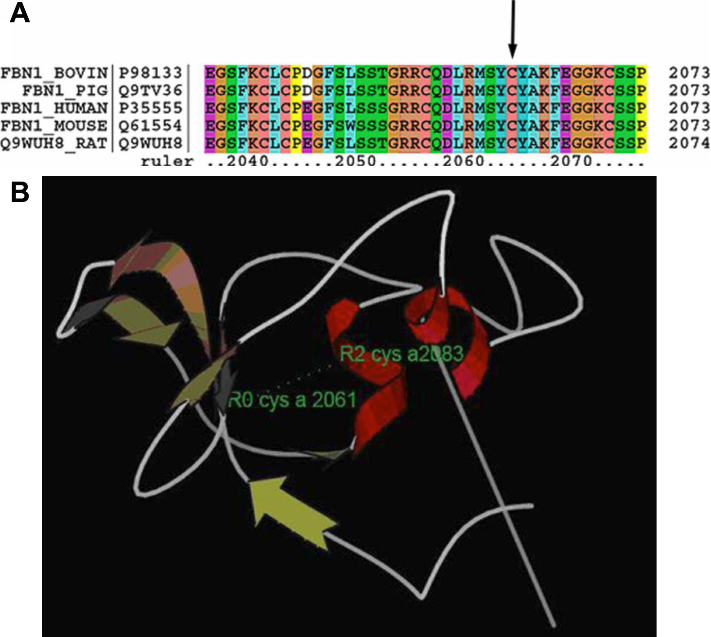
Figure 4. Analysis of the missense
mutation in exon 50.
A: The alignment of the FBN1 sequence with
the corresponding segments in diverse species is shown. The cysteine is
conserved in FBN1 proteins from several species. The sequence was
selected from the
UniProt
Knowledge base.
B: Structure analysis of the transforming
growth factor-binding protein-like domains (8-Cys/TB) in human FBN1.
α-helices and β-strands are shown with red and brown colors. The two
residues (C2061 and C2083) are colored green. The disulfide bond is
represented with a dotted line.
 Figure 4 of Zhao, Mol Vis 2009; 15:826-832.