Figure 4. Pro370Leu mutant myocilin induces ROS generation and increases calcium levels in the cytoplasm and mitochondria in TM cells
as estimated using flow cytometry. A: The oxidation sensitive fluorescence dye, H2-DCF-DA, was used to measure the ROS levels in TM cells. TM cells transfected with Pro370Leu (P370L) mutant myocilin transfection
show 2.05 fold (±0.94) stronger H2-DCF-DA fluorescent intensity than those transfected with WT myocilin (WT), indicating an increase in ROS generation after
Pro370Leu mutant myocilin transfection compared to WT myocilin transfection. B,C: The [Ca2+]c and [Ca2+]m indicators, fluo-3/AM and Rhod-2, were used to illustrated the free Ca2+ levels in the cytoplasm and mitochondria, respectively. There was a 3.17 fold (±0.64) increase in [Ca2+]c and 1.92 fold (±0.74) increase in [Ca2+]m after Pro370Leu mutant myocilin (P370L) transfection compared to WT myocilin transfection (WT). Data are expressed as fold
changes in fluorescent levels of transfected TM cells to non-transfected TM cells (Control). Results are expressed as the
mean±SE of three repeated experiments done in triplicate. An asterisk indicates a significant difference from non-transfected
cells at p<0.05.
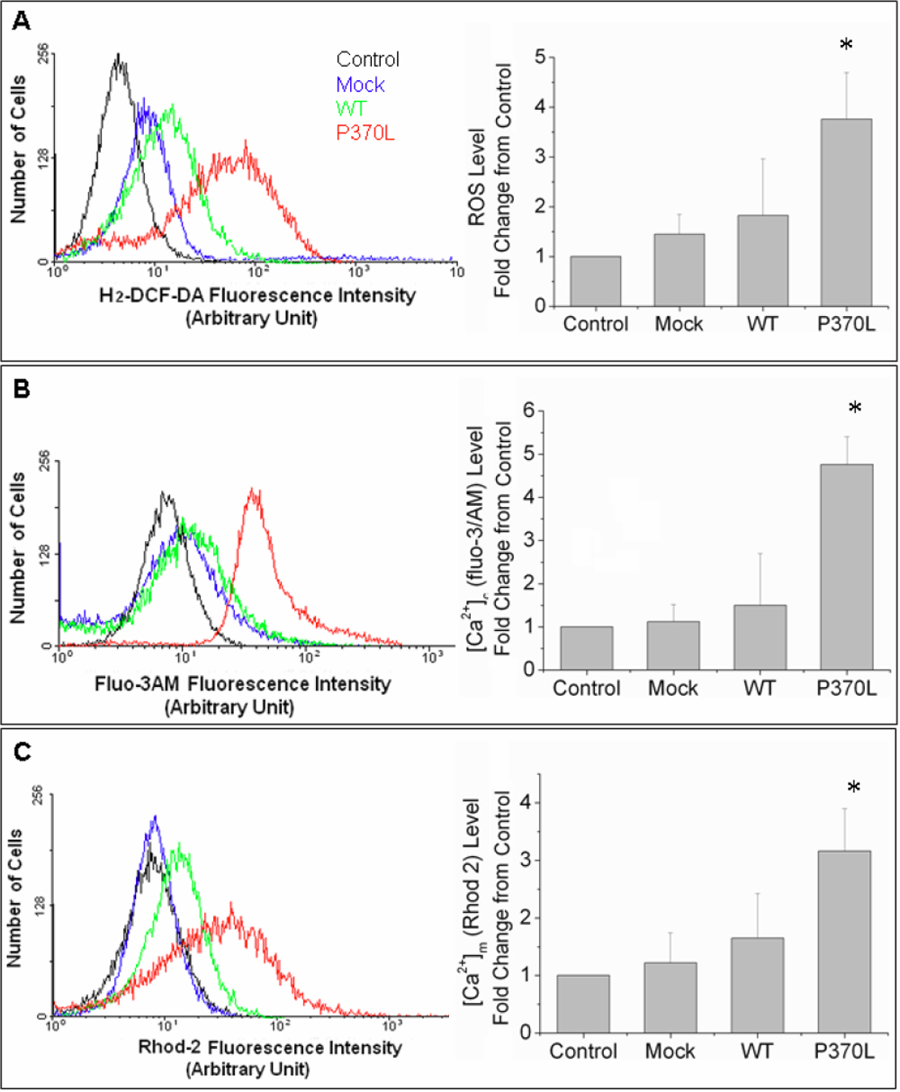
 Figure 4 of
He, Mol Vis 2009; 15:815-825.
Figure 4 of
He, Mol Vis 2009; 15:815-825.