Figure 3. Increased expression of myocilin after transfection. RT- PCR and western blot show that transfection of WT or Pro370Leu (P370L)
mutant myocilin increase both myocilin mRNA and protein levels in TM cells after 48 h. GAPDH was used as the internal loading control in RT–PCR while β-actin and cytochrome C oxidase (COX IV) were used as internal
loading controls in western blot analysis for total cell lysate and mitochondrial lysate, respectively. Confocal images show
myocilin co-localized with mitochondria. After transfection for 8 h, non-fixed TM cells were immunolabeled with MitoTracker
Red, a mitochondria specific dye. Confocal microscopic analyses show that both pIRES-EGFP-WT myocilin (WT) and pIRES-EGFP-Pro370Leu
mutant myocilin (P370L) transfected cells have overlapping EGFP staining (green fluorescence) and mitochondria staining (red
fluorescence). However, the green fluorescence from pIRES-EGFP (Mock) is diffusive within the cells with no overlapping with
mitochondrial staining. Our result indicates co-localization of myocilin and mitochondria in TM cells. Scale bar=30 μm.
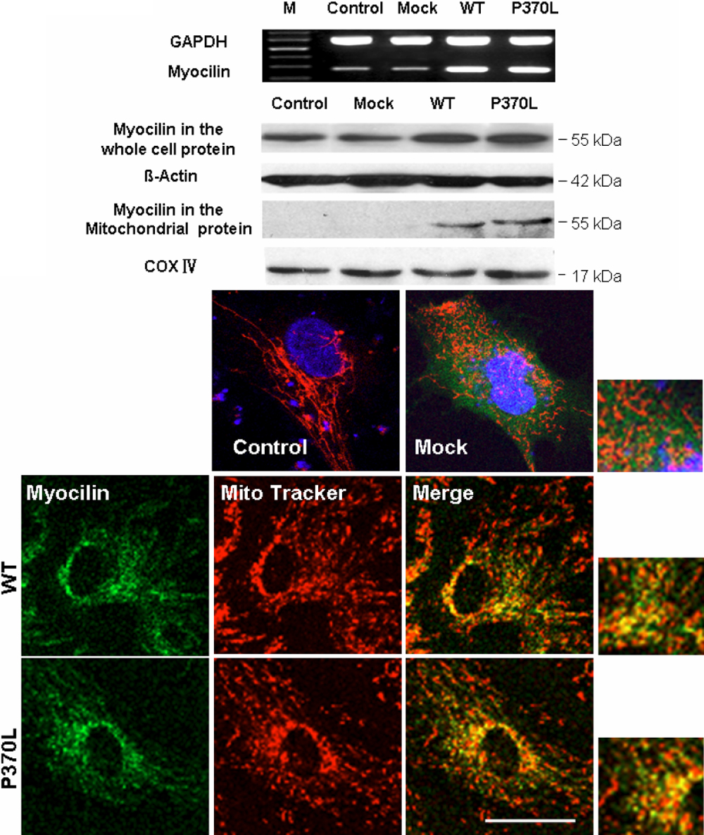
 Figure 3 of
He, Mol Vis 2009; 15:815-825.
Figure 3 of
He, Mol Vis 2009; 15:815-825.