Figure 1. Morphology change of TM cells
after Pro370Leu mutant myocilin transfection. Phase-contrast light
micrographs show healthy looking TM cells in non-transfected (Control),
pIRES transfected (Mock), or pIRES-WT myocilin transfected (WT)
cultures. However, TM cells demonstrate an unhealthy, degenerative
appearance after transfection with pIRES-Pro370Leu mutant myocilin
(P370L) for 24 h. Fewer cells are seen in these cultures in part due to
cell death and cell detachment. Scale bar=30 μm.
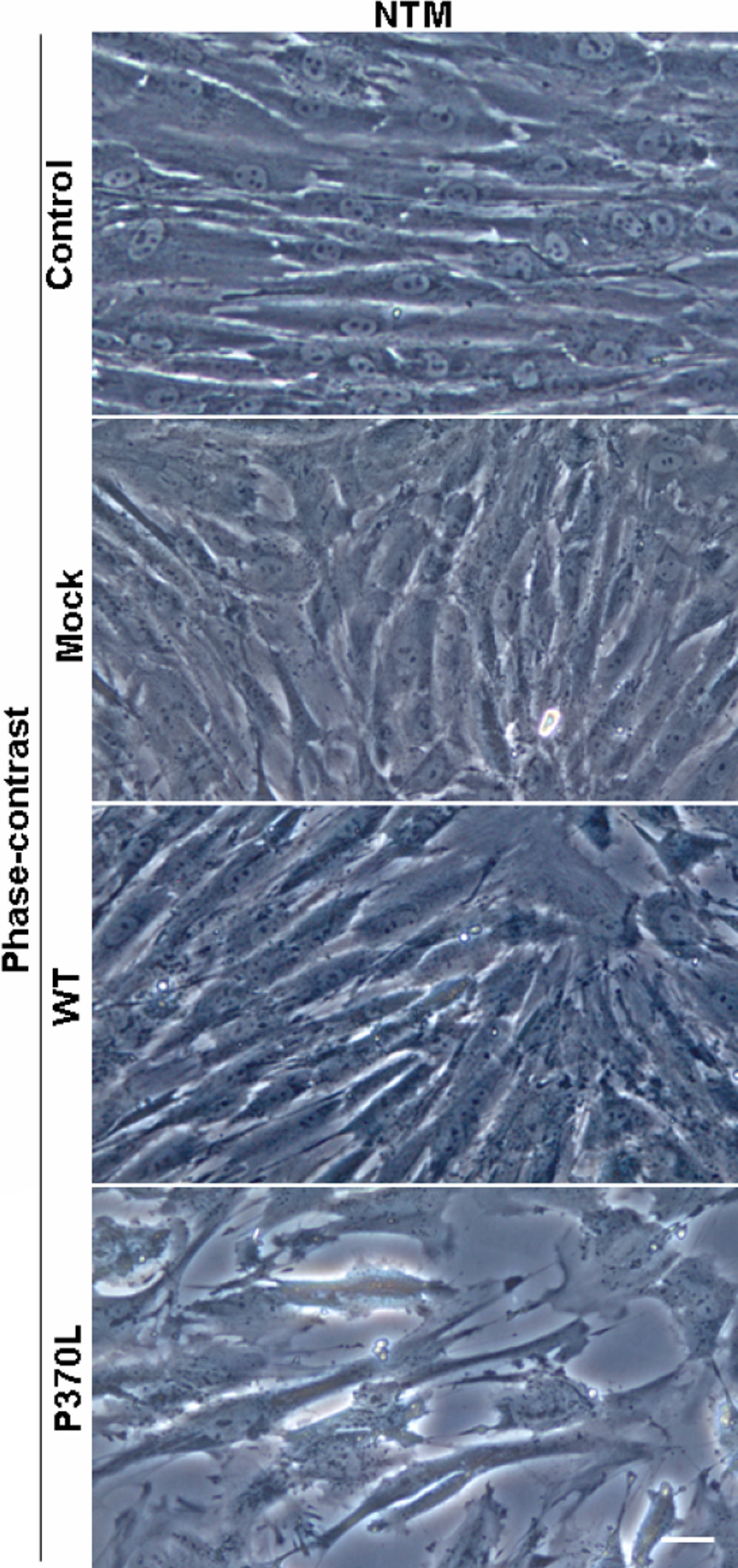
 Figure 1 of He, Mol Vis 2009; 15:815-825.