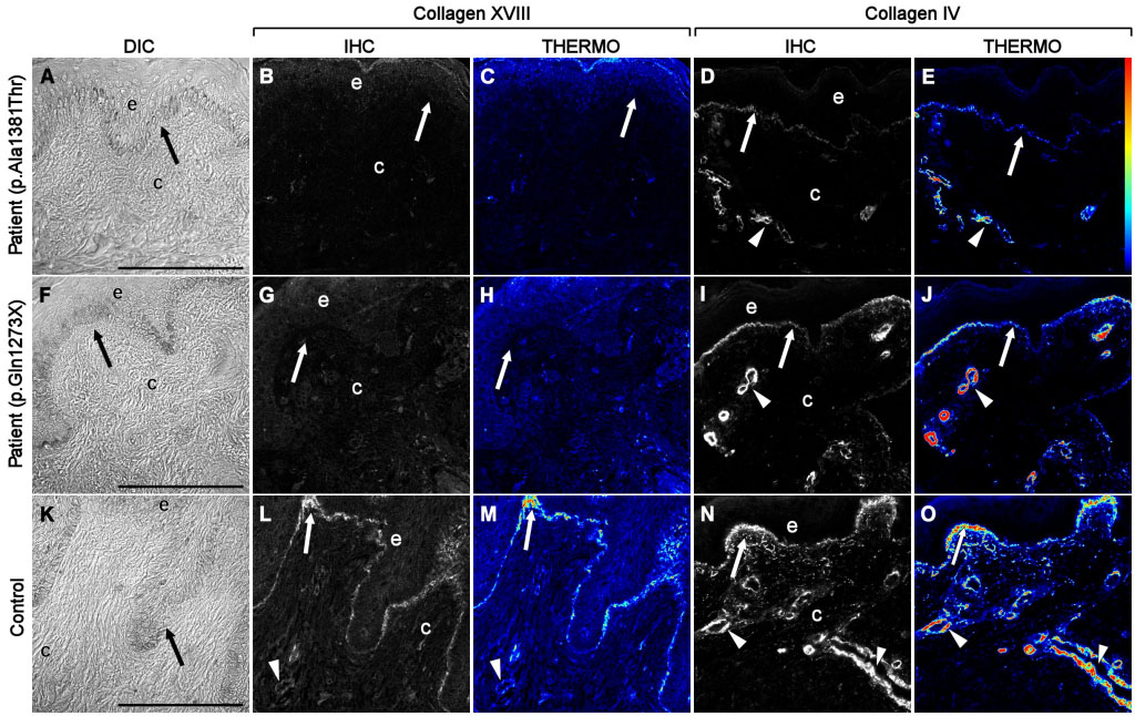
Figure 3. Immunofluorescent localization of type XVIII collagen in KS and control skin samples. Differential interference contrast (DIC)
images (
A, F, K) show that the epithelial and connective tissues obtained from skin biopsies were intact and well preserved after being prepared
using cryofixation and cryosubstitution techniques. Immunolocalization analyses of KS patients carrying the p.A1381T change
(
B, C) and the nonsense mutation c.3277C>T (NC11–493 isoform position p.G1273X,
GenBank AF018081.1;
G, H) were negative for expression of collagen XVIII as compared to controls (
L, M). Pseudocolor, thermo images showed that collagen XVIII expression was present in control samples (
M) but undetectable in KS samples (
C, H). Although the distribution pattern of type IV collagen was similar in both control (
N, O), and KS (
D, E, I, J) groups, the immunostaining intensity was noticeably lower in KS samples. The thermo color bar to the right of panel
E indicates immunofluorescence staining intensity: red, higher levels; blue/black, lower levels. Arrows point to basement membrane,
and arrowheads mark blood vessels. Scale bars equal 100 μm.
 Figure 3 of
Suzuki, Mol Vis 2009; 15:801-809.
Figure 3 of
Suzuki, Mol Vis 2009; 15:801-809.