Figure 2. Mutation analysis of CRYGD in Family 10. A:
Pedigree of Family 10 shows the proband, which is indicated by the
arrow. B: Direct sequencing of the PCR product encompasses exon
2 of CRYGD of an unaffected individual (I-2). C: Direct
sequencing of the PCR product encompassing exon 2 of CRYGD
shows a heterozygous TAC>TAG transition that replaced a tyrosine by
a premature stop codon at amino acid 56 (Y56X) in individual II-1. D:
The photograph of the anterior eye with lens image of individual II-1
shows nuclear cataract. E: Multiple alignment of amino acid
sequence of γD-crystallin protein with different species is shown:
mouse (1), rat (2), human (3), cow (4), and kangaroo (5). The Y56
residue is marked in red.
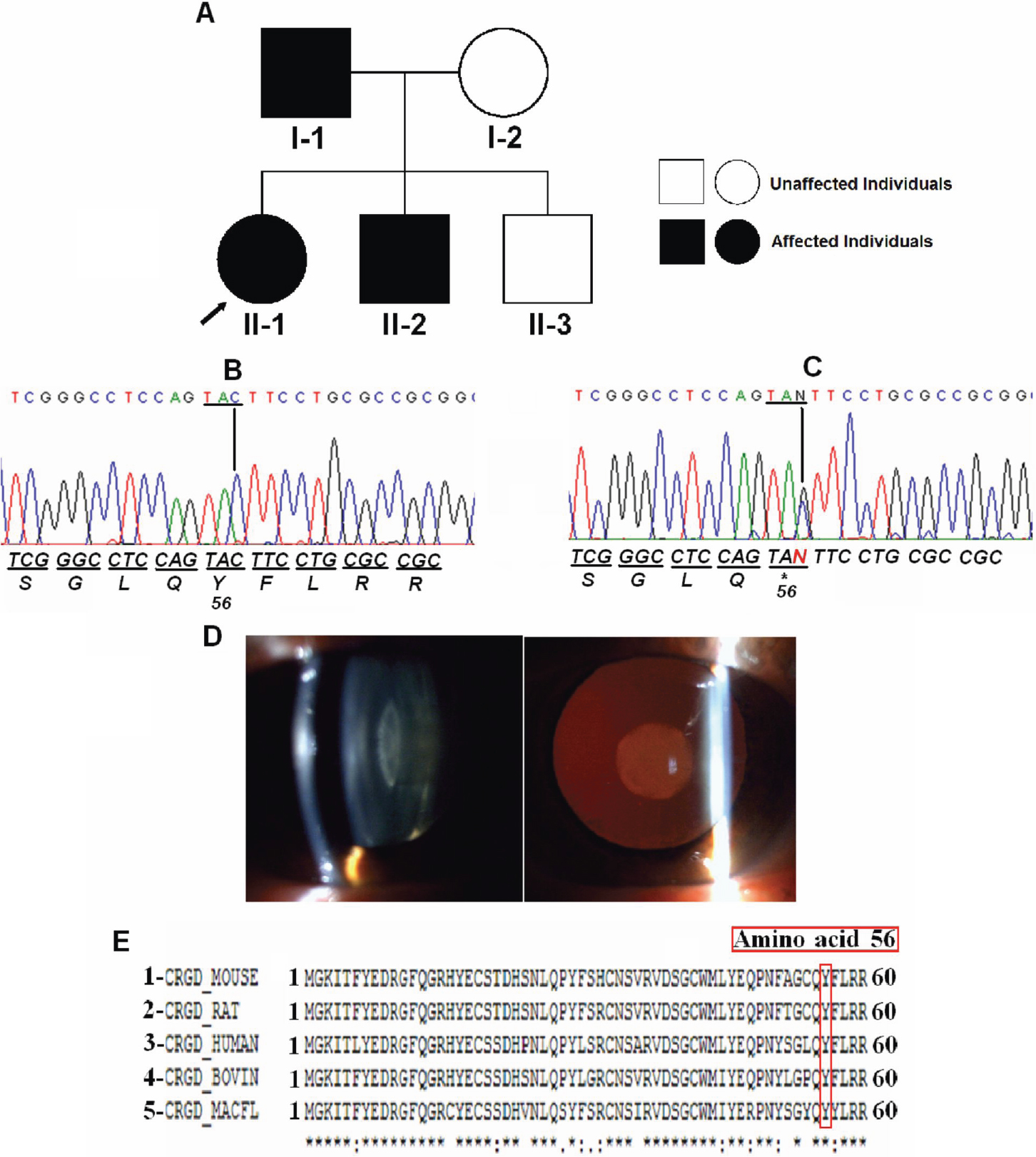
 Figure 2 of Santana, Mol Vis 2009; 15:793-800.