Figure 1. Mutation analysis of CRYAA in Family 4. A:
Pedigree of Family 4 shows the proband, which is indicated by the
arrow. B: Direct sequencing of the PCR product encompasses exon
1 of CRYAA (5′→3′) of an unaffected individual (I-1). C:
Direct sequencing of the PCR product encompassing exon 1 of CRYAA
of an affected individual (II-1) shows a heterozygous C→T transition
that replaced arginine by cysteine at amino acid 12 (R12C). The mutated
sequence is shown in red. D: The slit-lamp photograph of
individual I-2 shows a nuclear cataract. E: Alignment of
residues 1–60 of human (8) αA-crystallin protein with rat (1), hamster
(2), mouse (3), guinea pig (4), cow (5), sheep (6), pig (7) is shown.
The R12 residue is marked in red.
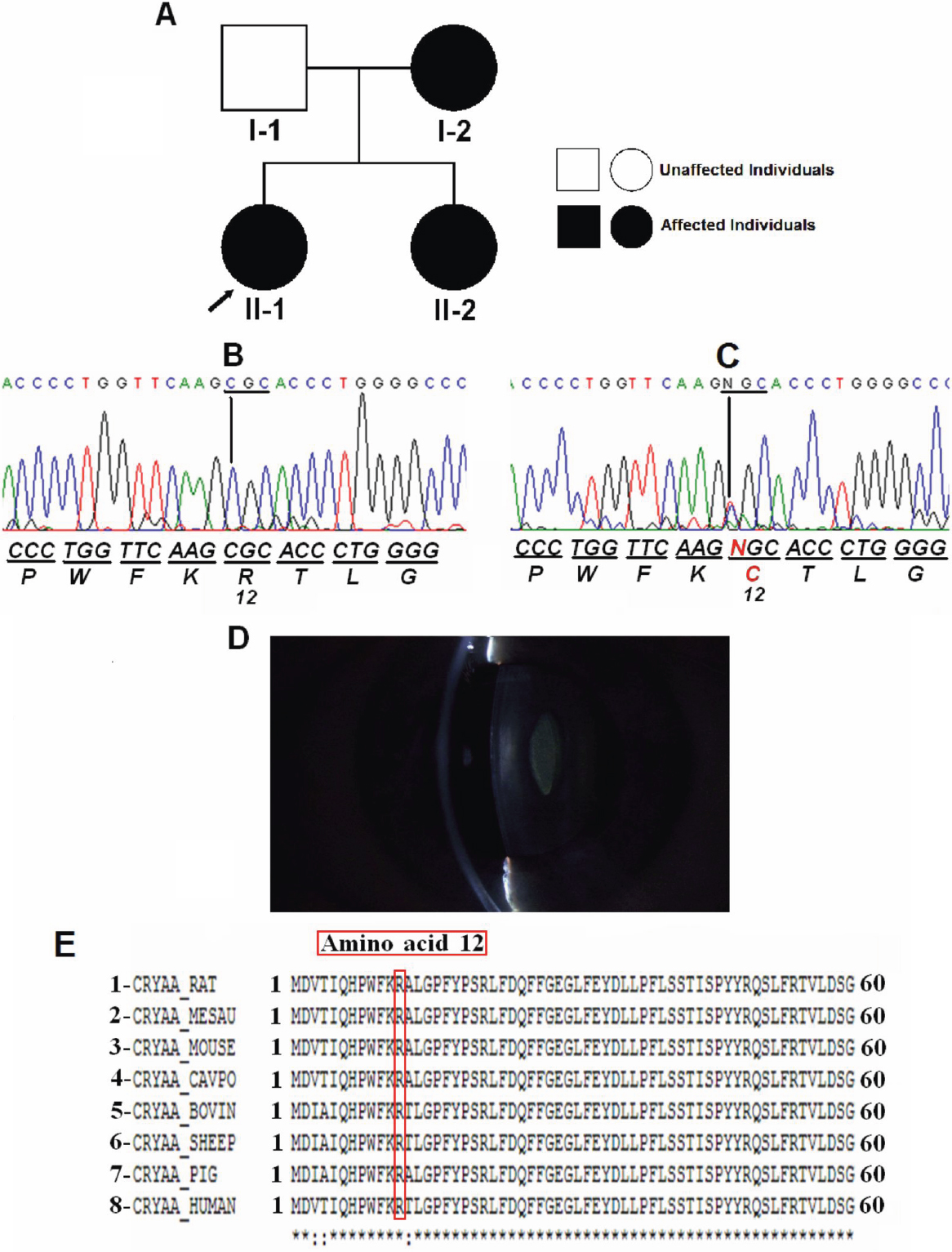
 Figure 1 of Santana, Mol Vis 2009; 15:793-800.