Figure 5. Distribution of ATH in the
sclera and choroid of control and recovering chick eyes. Monoclonal
antibodies specific for ATH were used together with AlexaFluor
488-conjugated rabbit anti-mouse immunoglobulin to localize ATH in
sclera and choroid of control and recovering eyes. A: Intense
ATH immunolabeling is detected in the fibrous sclera (FS) and in
perivascular and extravascular regions of the choroid. ATH was absent
in the cartilaginous scleral layer (CS). B: IgG control section
of the sclera and choroid of a control eye, where nonimmune mouse IgG
was used in the first incubation, followed by incubation in AlexaFluor
488-conjugated rabbit anti-mouse immunoglobulin. C: ATH
immunolabelling in the sclera of a recovering chick eye is shown in a
region artifactually separated from the choroid. ATH can be seen
localized in the outer fibrous sclera (FS) as well as in the thin inner
fibrous sclera on the choroidal side of the sclera. D: ATH
immunolabeling in the choroid of a recovering chick eye is shown. ATH
can be seen throughout the stroma of the markedly expanded choroid and
on the choroidal side of Bruch’s membrane (arrow), but is absent in the
RPE. Nuclei were stained with DAPI (blue). Choriocapillaris is
abbreviated CC. Scale bar (A–D) represents 50 μm.
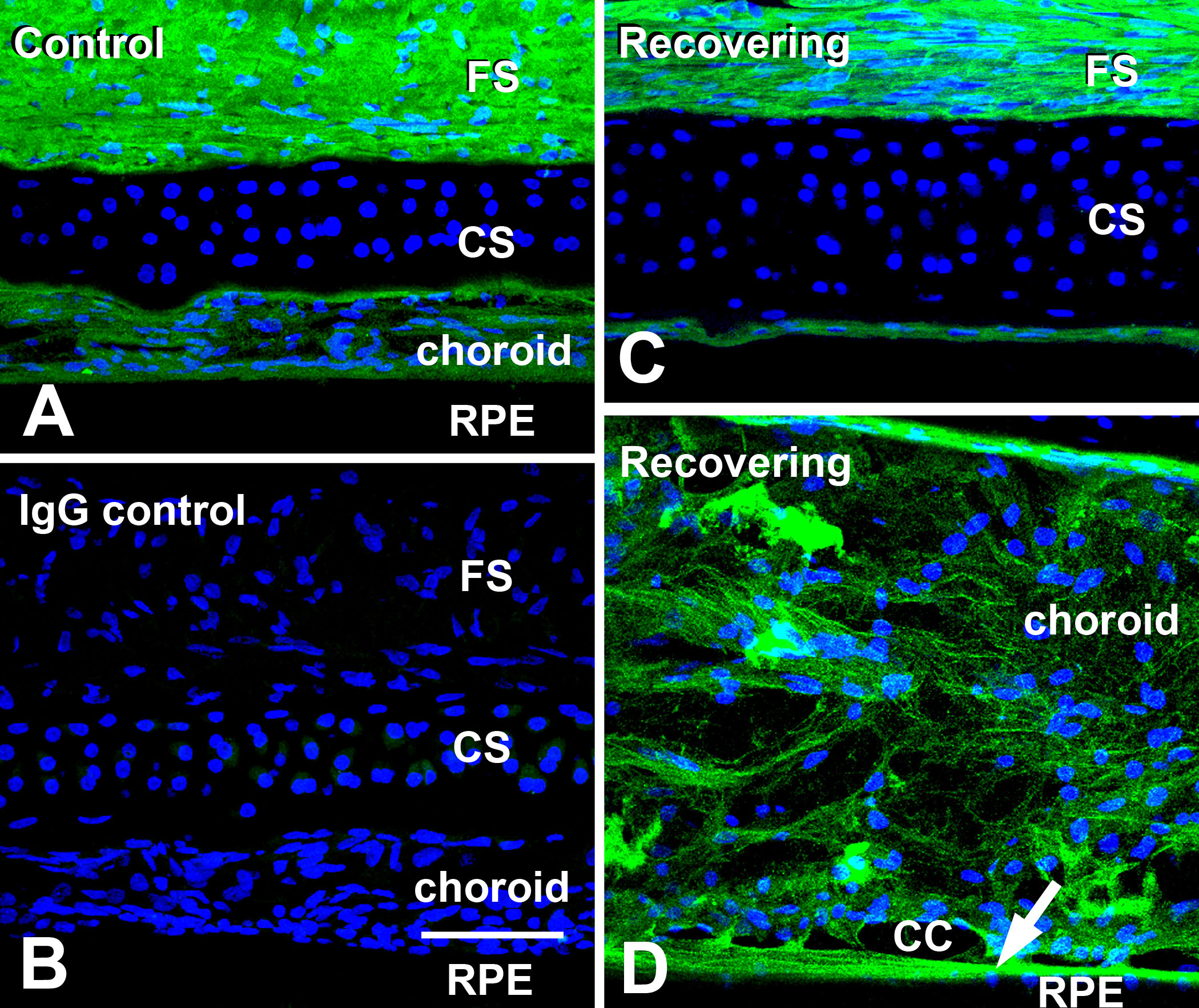
 Figure 5 of Rada, Mol Vis 2009; 15:778-792.