Figure 4. Distribution of ATH in the retina of control and recovering chick eyes. A: ATH immunolabeling in the retina of a control chick eye ATH immunoreactivity can be seen in a subpopulation of amacrine cells
in the inner nuclear layer (INL) as well as in proximal and distal sublaminae in the inner plexiform layer (IPL; arrowheads).
Nonspecific labeling is present in lipid droplets in the photoreceptor inner segments and in the nerve fiber layer (*). Inset:
A presumptive ATH-immunoreactive bistratified amacrine cell is demonstrated with a process appearing to extend to the distal
IPL (arrowhead). Scale bar represents 50 μm. B: ATH immunolabeling is shown in the retina of a recovering chick eye. Similar to the control eye, ATH immunoreactivity can
be seen in a subpopulation of amacrine cells in the INL as well as in proximal and distal sublaminae in the IPL (arrowheads).
C: IgG control section of the retina of a control eye is shown, where non-immune mouse IgG was used in the first incubation,
followed by incubation in AlexaFluor 488-conjugated rabbit anti-mouse immunoglobulin. Scale bar (A–C) represents 50 μm.
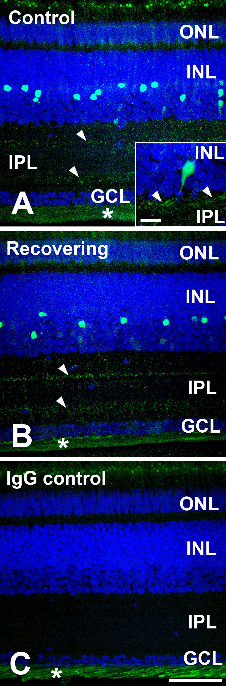
 Figure 4 of
Rada, Mol Vis 2009; 15:778-792.
Figure 4 of
Rada, Mol Vis 2009; 15:778-792.