Figure 3. Localization of Avian Thymic
Hormone (ATH) labeling in posterior ocular tissues of control and
recovering chick eyes. Monoclonal antibodies specific for ATH (obtained
from Michael Henzel, University of Missouri, Columbia, MO) were used
together with AlexaFluor 488-conjugated rabbit anti-mouse
immunoglobulin to localize ATH in chick ocular tissues. A: ATH
immunolabeling of a control eye demonstrated intense labeling in the
fibrous scleral layer (FS) and the choroid. Additionally, ATH was
detected in a subpopulation of cell bodies located in the inner nuclear
layer (INL) and in two sublaminae of the inner plexiform layer, most
likely representing the synaptic processes of ATH-positive cells of the
INL. B: IgG control section of a control eye is shown where
non-immune mouse IgG was used in the first incubation, followed by
incubation in AlexaFluor 488-conjugated rabbit anti-mouse
immunoglobulin. Fluorescence of lipid droplets in the photoreceptor
inner segments (*) and in the nerve fiber layer is nonspecific. Nuclei
were stained with DAPI (blue). C: ATH distribution in a
recovering eye was similar to that of controls, with specific
immunolabeling in the fibrous sclera, as well as throughout the
markedly thickened choroid, cells of the INL and sublaminae of the
inner plexiform layer (IPL). Vertical bars indicate thickness of
choroid layer in control (3A) and recovering (3C) eyes. Abbreviations:
cartilaginous sclera (CS), outer nuclear layer (ONL), ganglion cell
layer (GCL). Scale bar (A–C) equals 100 μm.
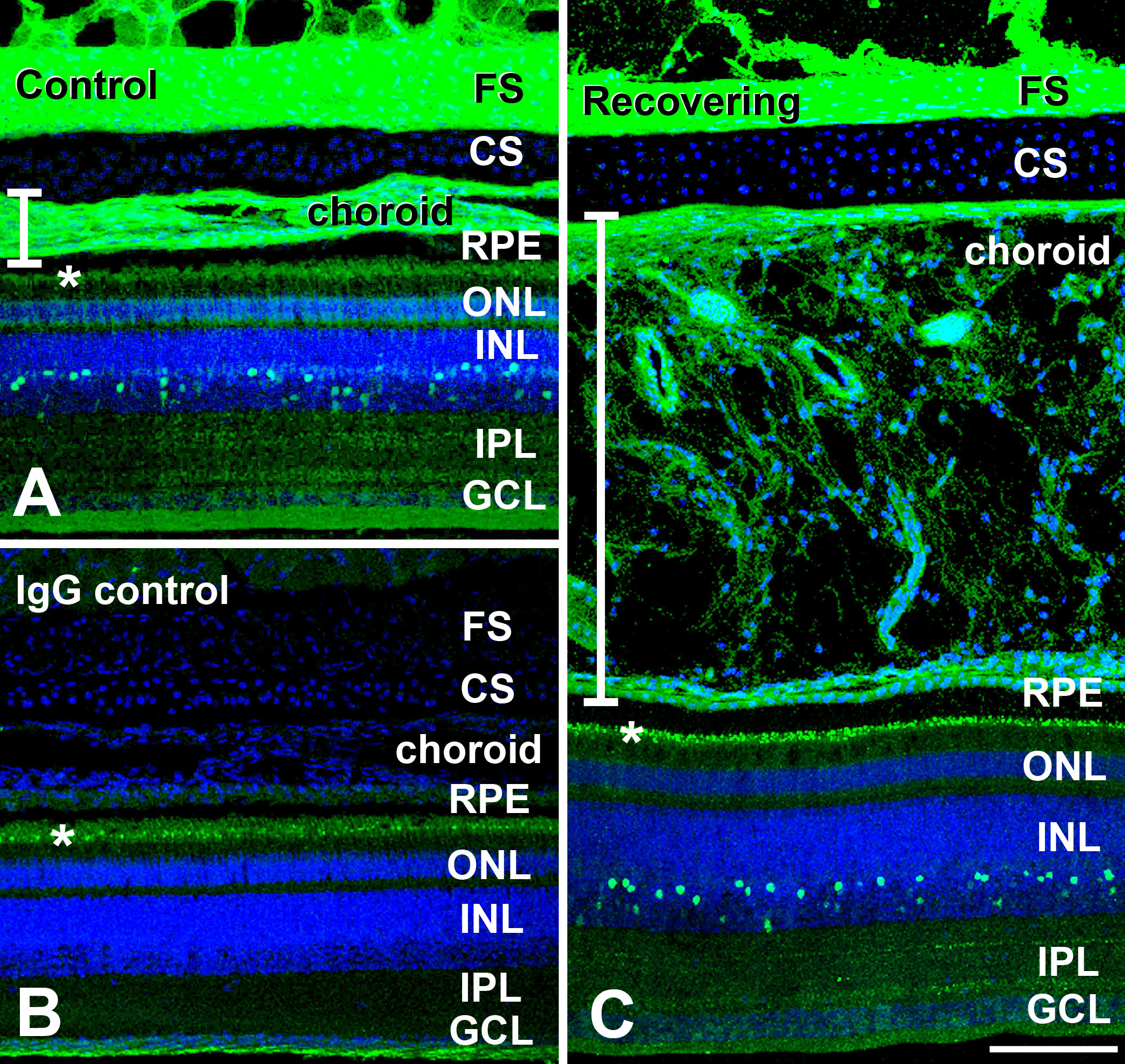
 Figure 3 of Rada, Mol Vis 2009; 15:778-792.