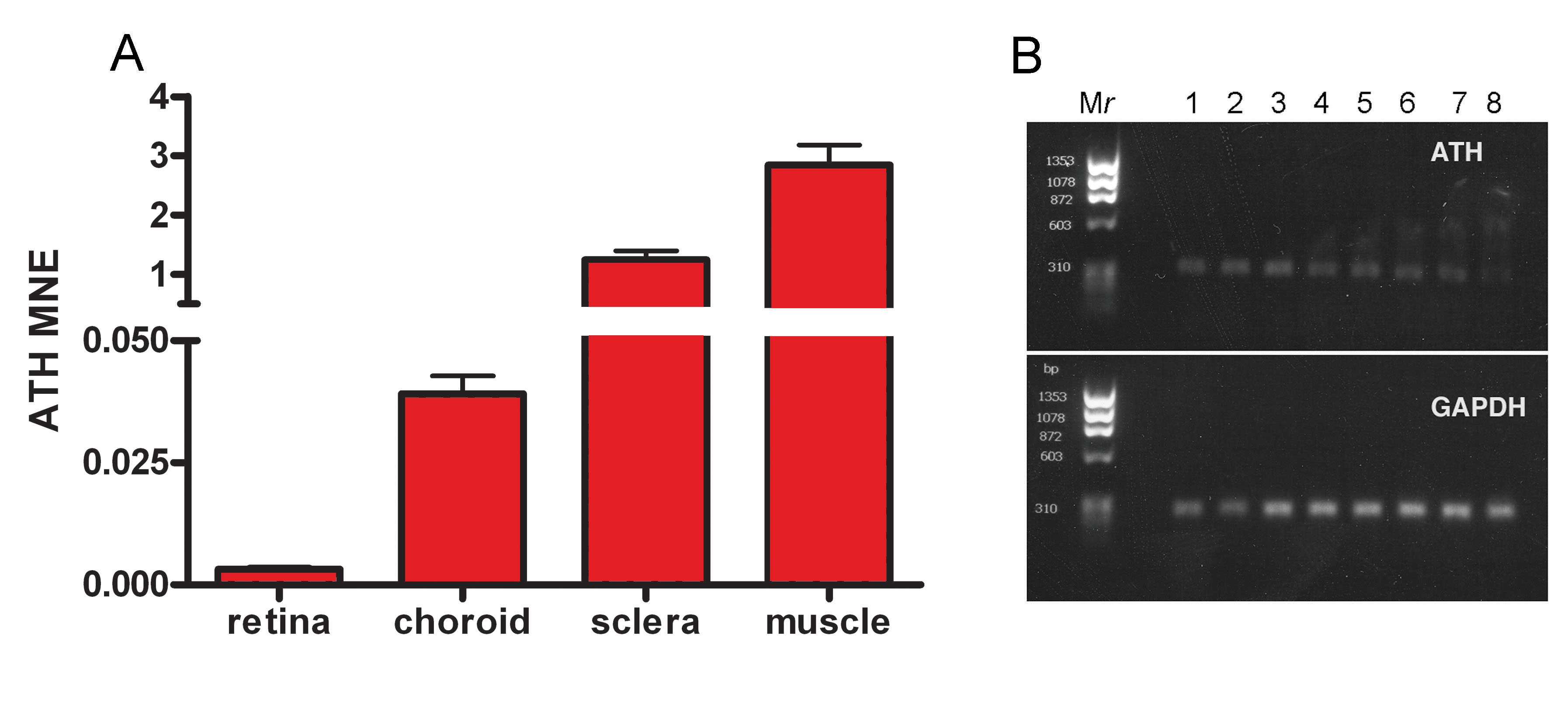
Figure 1. Real time RT–PCR quantification
of
ATH in chick ocular tissues. Steady-state levels of
ATH
mRNA were measured in the retina–RPE, choroid, sclera, and extraocular
muscle using chick gene-specific primers (
Table 1).
A: The average
mean normalized expression (MNE) of ATH was calculated in each ocular
tissue of three untreated 10 day old chicks. All reactions were run in
triplicate and normalized to the reference gene,
GAPDH.
B:
Shown are ethidium bromide gels of real-time PCR products of
ATH
and
GAPDH in chick extraocular muscle over temperatures ranging
66–55 °C. M
r: PhiX174RF DNA/HaeIII molecular weight ladder,
Lane 1: 66.0 °C, Lane 2: 65.2 °C, Lane 3: 63.9 °C, Lane
4: 61.8 °C, Lane 5: 59.0 °C, Lane 6: 57.2 °C, Lane 7:
55.8 °C, Lane 8: 55.0 °C.
 Figure 1 of Rada, Mol Vis 2009; 15:778-792.