Figure 4. Representative immunofluorescence histochemistry of normal and glaucomatous human optic nerve head lamina cribrosa tissue.
Normal (A) and glaucomatous (B) optic nerve head sections were stained for Periostin (green). Periostin was increased in the lamina cribrosa of glaucomatous
sections compared to the normal controls. No immunostaining was seen in the absence of primary antibody (C and D shows blue DAPI staining of nuclei).
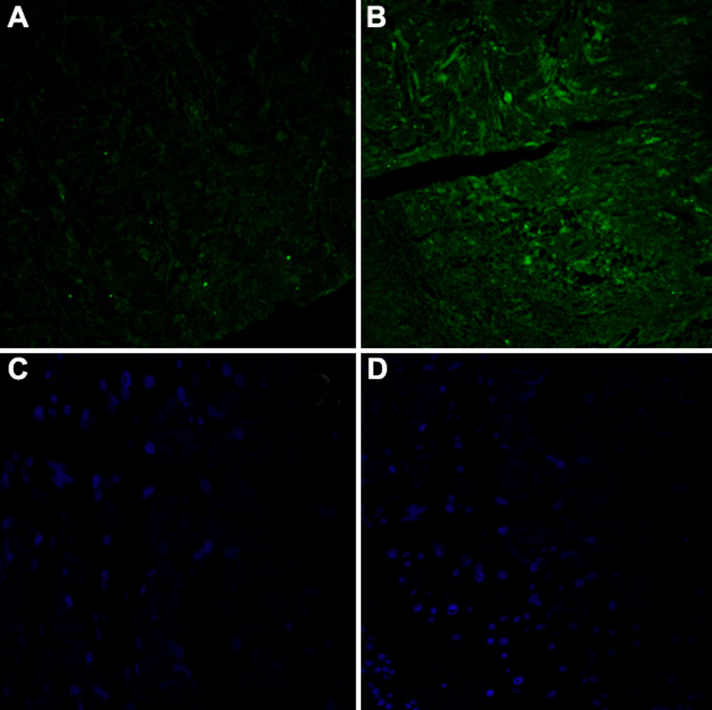
 Figure 4 of
Kirwan, Mol Vis 2009; 15:76-88.
Figure 4 of
Kirwan, Mol Vis 2009; 15:76-88.